In my last post, I started a series on Cranial Nerves, in other to put clarity to the functions the cranial nerves have on our sense organ, and how they take this sense signals to the brain. In my last post Neurology Explained - The Olfactory nerve (Cranial Nerve 1) I explained the first Cranial Nerve which was the Olfactory nerve. It is responsible for picking smell particles and taking the smell to the brain, which interprets and give the rewards to the smell. In my post today, I will be looking into the Cranial Nerve II, which is the Optic Nerve.
To understand how the optic cranial nerve works, you need to first understand the phototransduction process of the eye. I explained how light ray is converted into chemicals then electrical signals, which are sent to the brain. You remember the Cones and Rods in my post Explaining Phototransduction in the Eye.
After the Retinal pigment epithelium are the photoreceptors, which are very important in Phototransduction. It is made up of the Rods and the Cones. The Rods are specifically meant for scotopic visions (dark/dim light). It is required during night vision. It is a cylindrical structure of plasma membrane, made up of pigment known as Rhodopsin which is a combination of Retinal (11-cis retinal) and opsin. Cones on the other hand are responsible in photopic vision. Cone is made up of a pigment known as the cone opsin called photopsin which is made up of photopsin 1, photopsin 2, and photopsin 3, iodine, and opsin with cone visual pigments, allowing the eye to be able to pick up color from different wavelengths. - Explaining Phototransduction in the Eye
When your eyes look at an object, the eye does the balancing, and it is able to see other things and background, in a fixed point. The region where clear sight is visible by the is regarded as Visual field. The Visual field of the eyes would be the right and the left eye visual field. The visual fields are further classified by their ability of sight and border. The one close to the nose is the Nasal Visual Field Border of the eye, while the one away from the nose and towards the temporal region (between the eye and the ear) is regarded as the Temporal Visual Field Border of the eye..
The Temporal Retina receives light rays from the nasal visual field border, and the Nasal Retina receives light ray/visual information from the temporal visual field border. The Right eye, has the Temporal Retina, and the Nasal Retina, also does the Left eye have the Temporal Retina and the Nasal Retina. The Temporal Retina of the right eye receives light rays from the left visual field of the left eye, and the Nasal Retina of the left eye receives light rays from the right visual field of the left eye. On the Left eye, the Temporal retina receives light rays from the right visual field of the right eye, and the Nasal retina receives light rays from the left visual field of the left eye..
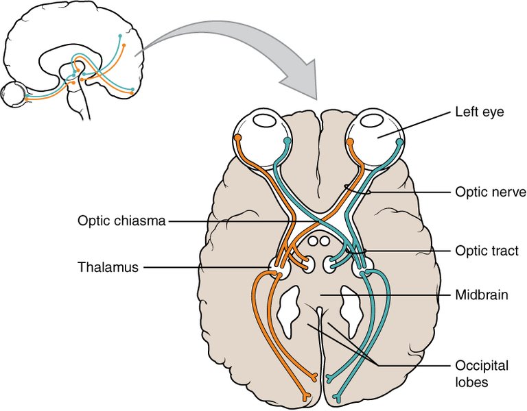
When the information is received by the Retina of both eyes, it is taken by the Optic nerves. Each eyeball has its optic nerves, and the optic nerves meet at a point close to the pituitary gland in the brain, called the Optic Chiasma.
Images from the Retina (Retina ganglion cells) moves through the optic tract (the right optic tract and the left optic tract), and reaches the dorsal thalamus to the lateral geniculate nucleus (LGN).. The lateral geniculate nucleus is made up of six layers, where the Ipsilateral and contralateral optic fiber are joined. The Ipsilateral fiber nerve connects to the 2nd, 3rd, and 5th layers of the lateral geniculate nucleus, and the Contralateral fiber nerve connects to the 1st, 4th, and the 6th layer of the lateral geniculate nucleus., .
From the lateral geniculate nucleus, some of the optic fiber go into Superior colliculus and the pretectal nucleus of the midbrain. The Superior colliculus is responsible for the eye movement when the head is restricted and the eye needs to move. The Pretectal nucleus of the eye is responsible for the control of the pupillary light reflex (the constriction of the pupil). Some optic fiber which are the (Meyer’s loop (Inferior retinal fibers)) from the lateral geniculate nucleus go through the temporal lobe on one end to the striate cortex of the Occipital lobe. Also, on another end from the lateral geniculate nucleus are the (Baum’s loop (Superior retinal fibers)) which moves from the perietal lobe on another end to the striate cortex of the Occipital lobe, which is the primary visual cortex.. The striate cortex is responsible for processing visual information gotten from the eye. It should be stated that Optic Nerve isn't the only cranial nerve responsible for innervating sight, other cranial nerves include the Cranial nerve III, Cranial nerve IV, Cranial nerve V, Cranial nerve VI, and the Cranial Nerve VII (I will be explaining these cranial nerves in subsequent posts. These Cranial Nerves innervate other functions beyond sight).
Conclusion
The Cranial Nerve II is a special sensory afferent nerve fiber that originates from the retina, as light rays are received by the Retina. The nerve picks information from the photoreceptors (Rods, Cones and the ganglion cells), to allow for visions. It is important to know that the Cranial Nerve II is a sensory nerve.
Well detailed information you presented in your article. Glad I was able to pick some new knowledge
I am happy that you were able to pick some little knowledge. I am really glad to have you on my blog. I hope to see more of you. Thanks
Thanks for your contribution to the STEMsocial community. Feel free to join us on discord to get to know the rest of us!
Please consider delegating to the @stemsocial account (85% of the curation rewards are returned).
Thanks for including @stemsocial as a beneficiary, which gives you stronger support.
I really like this series of posts! They re very clear and instructive (sorry, no question or comment once again). Thanks for choosing STEMsocial to share them with the world!
Thanks a lot @lemouth for always visiting my blog, you really encourage me to keep creating these contents. I am always glad to have you here. Thanks a bunch😘😘😘
Hehe! You are welcome my friend ;)