I actually wrote "hello everyone" several times, but cleared it out at every point I did, because I wasn't certain if it was a good way to start the post. I am sure you would be expecting me to create a post on the 8th cranial nerve (the Vestibulocochlear Nerve), as a continuation of my cranial nerve series, the answer is Yes, I will be creating a post on the 8th cranial nerve, but it won't be today, as there are a few things I should share before creating a post on the Vestibulocochlear nerve. To better understand the Vestibulocochlear nerve, it is important to have the knowledge about the anatomy of the inner ear, the cochlear, the vestibule, and the semicircular canal. I have been able to do justice to the anatomy and illustration of the inner ear, you can read about it in the post The Anatomy, and Physiology of the Ear. Before I continue with the 8th cranial nerve (the Vestibulocochlear Nerve) I will create a post on the cochlear, the vestibule, and the semicircular canal, so I will be starting with the Cochlear.
The Anatomy of the Cochlea
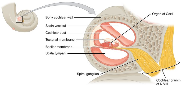
In the inner ear, is a snail-like structure known as the cochlea, which has outer bony part, with its upper part known as the Modiolus, a superior duct filled with perilymph which is high is sodium and low in potasium known as the Scala vestibuli or vestibular duct which is the end of the oval window, which has a lower duct known as the Scala tympani which is at the end of the round window, and in the middle of both the scala vestibula at the upper part in the cochlea, and the Scala Tympani, at the lower part of the cochlea, is the Cochlear duct which is made up of endolymph (fluid with high potassium ion concentration and low sodium ion) in its cavity. Extending from the Modiolus, are bony projections known as the Spiral Limbus, which is situated on the osseous spiral lamina.,
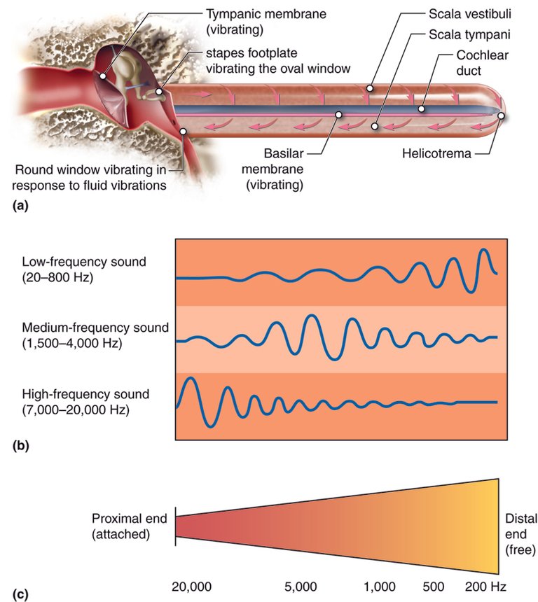
When sound waves enter into the ear, they hit the tympanic membrane, which changes the sound wave to mechanical energy, causing the tympanic membrane or eardrum to vibrate which the ear ossicles (the malleus, incus, and stapes). The Stapes is the last ossicle to be moved, and the stapes vibrates and hits the oval window, converting the mechanical energy into fluid filled vibrations, to the scala vestibuli. The Scala Vestibuli is seperated from the Scala media/cochlear duct by a membrane known as the reissner's membrane, also the Scala Tempani is separated from the Scala media by the basilar membrane, which is responsible for identifying different sound frequency (pitches).
In the Basilar Membrane, high-frequency sound waves stimulates the basilar membrane cochlear base, as a result of stiff and and short fibers towards the base of the membrane, and as they go towards the tip of membrane reaching the Helicotrema, the membrane becomes longer and floppy. The apex area of the basilar membrane which has a long and floppy membrane is known to receive low pitches. As the membrane moves towards the apex, the frequency it receives reduces. If a sound wave doesn't create a vibration in the basilar membranes, sounds won't be heard, which means it is too low, and any sound extremely high won't be heard as well.
Reticular Lamina is a collagenous fiber below the basal lamina, and surrounds the rods of corti. Going through the recticular lamina are hair cells known as inner hair cells and the outer hair cells. The inner hair cells serve as sensory receptors, which receives 95% of the peripheral processes, while only about 5 goes to the outer hair cells. Between the inner hair cells, and the outer hair cells is the spiral organ of corti.,
The inner hair cells have hair-like projections known as stereocilia which has its longest cilia being the kinocilium. The cilia are connected by proteins known as tiplinks. Above the inner hair cells is the Tectorial membrane, and remember that I said that the hair passes through the reticular lamina, and at the root is the basilar membrane, and between the inner hair cells and the outer hair cells are the rods of corti.. When the Basilar membrane vibrates, it makes the reticular lamina as well as the rod of corti to move upwards. As they move upwards, the reticular lamina causes the inner hair to be pulled inwards, causing the steriocilia to move towards the kinocilium allowing potassium and calcium to go into the hair cells, causing depolarization to release glutamate which acts on the spiral ganglion creating action potentials. ,
The Outer hair cells also have tiplinks and have proteins known as prestin which helps in the changing of the cell shape. When the prestin molecules contract, they cause the upward bowing of the basilar membrane thereby causing an increase in the hair cell activation thereby helping to discriminate different sounds. Extreme loud sounds, (high amplitude) activate the Olivocochlear bundle which then releases Acetylcholine which binds with outer hair cells, to allow potassium leave the cells to create hyper-polarized cell. Causing the cell to lengthen causing the basilar Bowing, where the basilar membrane goes downward, decreasing the vibration and of basilar membrane and at the same time decreasing the hair cells.,
Conclusion
When we receive sound from the ear, it hits the tympanic membrane (eardrum) which triggers the ossicles. The Ossicles are responsible for creating vibrations in the oval window which divides the cochlea from the middle ear. The Cochlea is a snail-shell shaped bony tube that is occupied by fluid (perilymph) in the upper part of the cochlea, and endolymph on the lower part of the cochlea. In the cochlea, the Scala Vestibuli, and the Scala Tympani is filled with the perilymph, while the Scala media is filled with the endolymph. The Basilar Membrane vibrates when there is wave movement in the cochlea. It is important to know that different part of the cochlea respond to different amplitude of sound. The inner hair cells and the outer cell hairs are part of the organ of corti. They help to maintain sound, regulate sound amplitudes and help to discriminate different sounds.
Image 1 ||Wikimedia Commons || File:1406 Cochlea.jpg
Image 2 || Coursehero.com || Special Senses: Hearing (Audition) and Balance
The rewards earned on this comment will go directly to the people( @academician ) sharing the post on Twitter as long as they are registered with @poshtoken. Sign up at https://hiveposh.com.
Thanks for your contribution to the STEMsocial community. Feel free to join us on discord to get to know the rest of us!
Please consider delegating to the @stemsocial account (85% of the curation rewards are returned).
Thanks for including @stemsocial as a beneficiary, which gives you stronger support.
This is a very detailed discussion on the cochlear. 👍