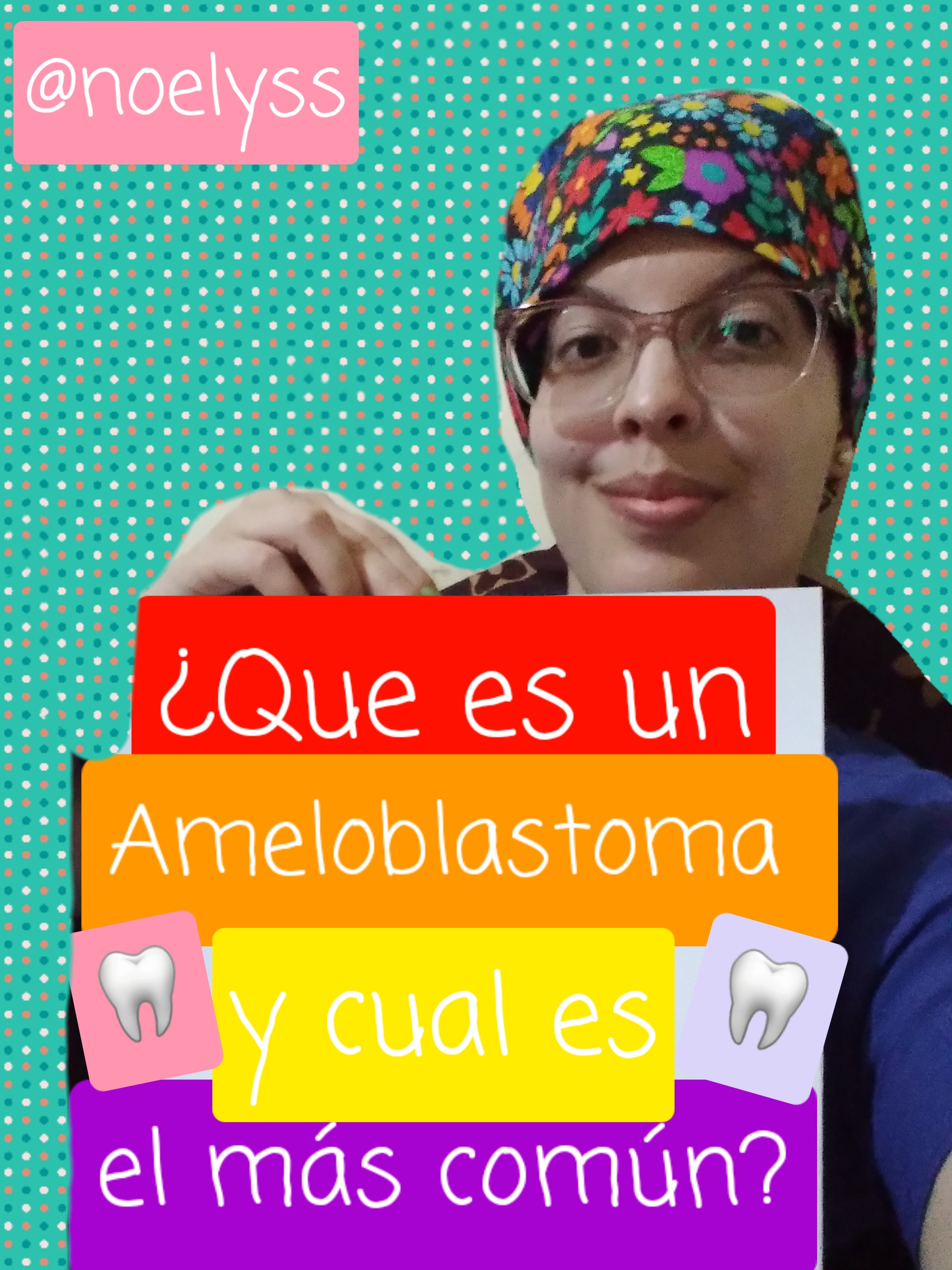
Seguimos profundizando acerca de los tumores y neoplasias ubicadas en la cavidad bucal, en esta oportunidad hablaremos del ameloblastoma qué es un tumor benigno este se forma debido al epitelio odontogénico, pero que tendrá un comportamiento algo invasor y con gran recidiva, qué será de una manera indiscriminada y agresiva por lo que ocasiona, en el paciente ciertas deformidades en el examen extraoral, debido a los restos de lámina dental de la mucosa que reviste la superficie.
We continue to deepen about tumours and neoplasms located in the oral cavity, this time we will talk about ameloblastoma, which is a benign tumour. opportunity to talk about ameloblastoma, which is a benign tumour that forms due to the odontogenic epithelium, but which will have a somewhat invasive and highly recurrent behaviour, which will be indiscriminate and aggressive, causing certain deformities in the patient in the extraoral examination. It tends to originate through the walls left by odontogenic cysts through the dental lamina of the mucosa that covers the surface.
Existen diferentes tipos de ameloblastoma los cuales se van a organizar por medio de una clasificación dónde encontraremos el ameloblastoma convencional que suele ser el más común de todos los ameloblastoma, es Sólido y multiquistico, también está el ameloblastoma uniquistico que este solamente lo vamos a encontrar en un 25% de los casos el ameloblastoma periférico que esté se encuentra fuera de los huesos y el ameloblastoma desmoplásico qué es sólido y posee características totalmente diferentes.
There are different types of ameloblastoma which are organised by means of a classification where we will find conventional ameloblastoma which is usually the most common of all ameloblastoma, it is solid and multi-cystic, there is also unicystic ameloblastoma which is only found in 25% of cases, peripheral ameloblastoma which is found outside the bones and desmoplastic ameloblastoma which is solid and has totally different characteristics.
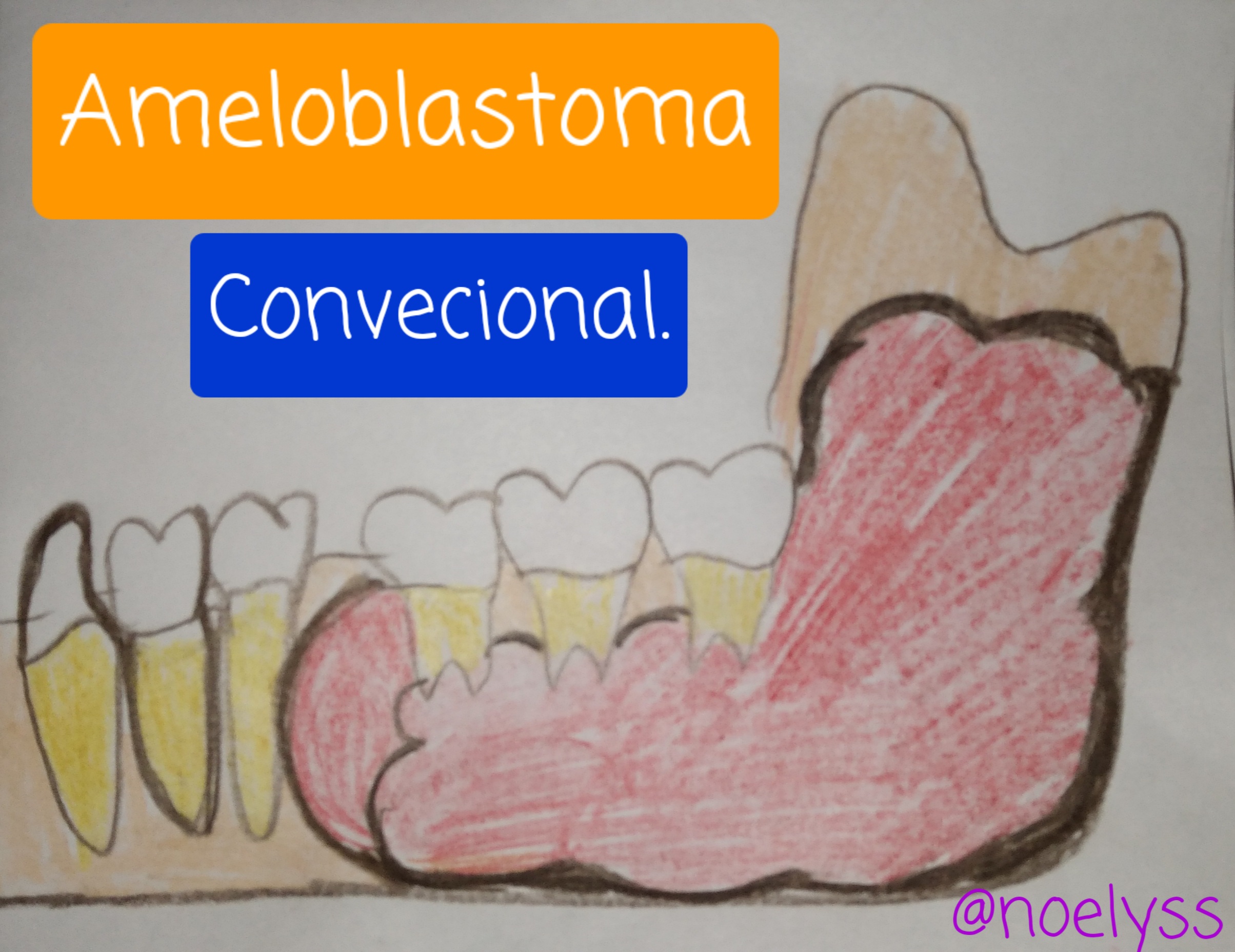
Ameloblastoma convencional el más común.
Conventional ameloblastoma is the most common.
Es el más común de todos los ameloblastoma No es maligno pero suele ser muy imperativo este tendrá consistencia sólida sin cavidades es multiquistico en cuánto hace la contra haremos en la primera década de la vida será poco frecuente en la segunda década de la vida pero no cante tendrá mayor predilección a los 30 años no tiene preferencia en cuanto al sexo ambos se pueden ver afectados, lo podemos localizar en la mandíbula específicamente en la zona posterior al nivel de los molares y de la rama ascendente al igual que el queratoquiste odontogénico.
It is the most common of all the ameloblastoma It is not malignant but it is usually very imperative this will have solid consistency without cavities is multiquistico in how much does the counter we will do in the first decade of life will be rare in the second decade of life but not sing will have greater predilection to the 30 years has no preference as to sex both can be affected, we can locate it in the jaw specifically in the posterior area at the level of the molars and the ascending branch as well as the odontogenic keratocyst.
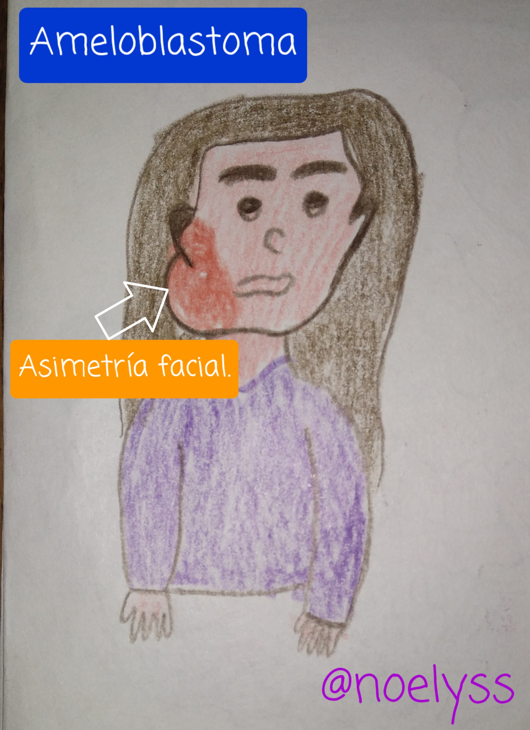
El paciente nos prepara sintomatología pero sí crecerá de manera desproporcionada, solo Existirá dolor cuando esté causa presión en algún nervio muy lento pero también puede ser de manera ilimitada es sólido Una vez que se extrae pero puede tener múltiples cavidades quísticas, para su tratamiento debemos proceder al abordaje quirúrgico mediante la cirugía Cabe destacar que va a depender del tamaño que esté posea si el tamaño es menor de un centímetro se debe hacer curetaje marginal con una fresa quitando la pared osea.
Once it is extracted but may have multiple cystic cavities, for its treatment we must proceed to the surgical approach by surgery. It should be noted that it will depend on the size of the cystic cavity, if the size is less than one centimetre, marginal curettage must be performed with a burr removing the bone wall.
Su pronóstico es reservado cuánto este se localiza en la mandíbula ya que se debe quitar parte del maxilar inferior, y es totalmente malo cuando afecta el maxilar superior ya que no se puede extraer, pero es bueno cuándo es pequeño, debemos saber que tiene una alta recidiva es decir que puede volver a aparecer y esto va a depender del tipo de tratamiento, ya que en el curetaje va a tener una alta recidiva.
Its prognosis is reserved when it is located in the mandible as part of the lower jaw must be removed, and it is totally bad when it affects the upper jaw as it cannot be extracted, but it is good when it is small, we must know that it has a high recurrence, that is to say that it can reappear and this will depend on the type of treatment, as in curettage it will have a high recurrence.
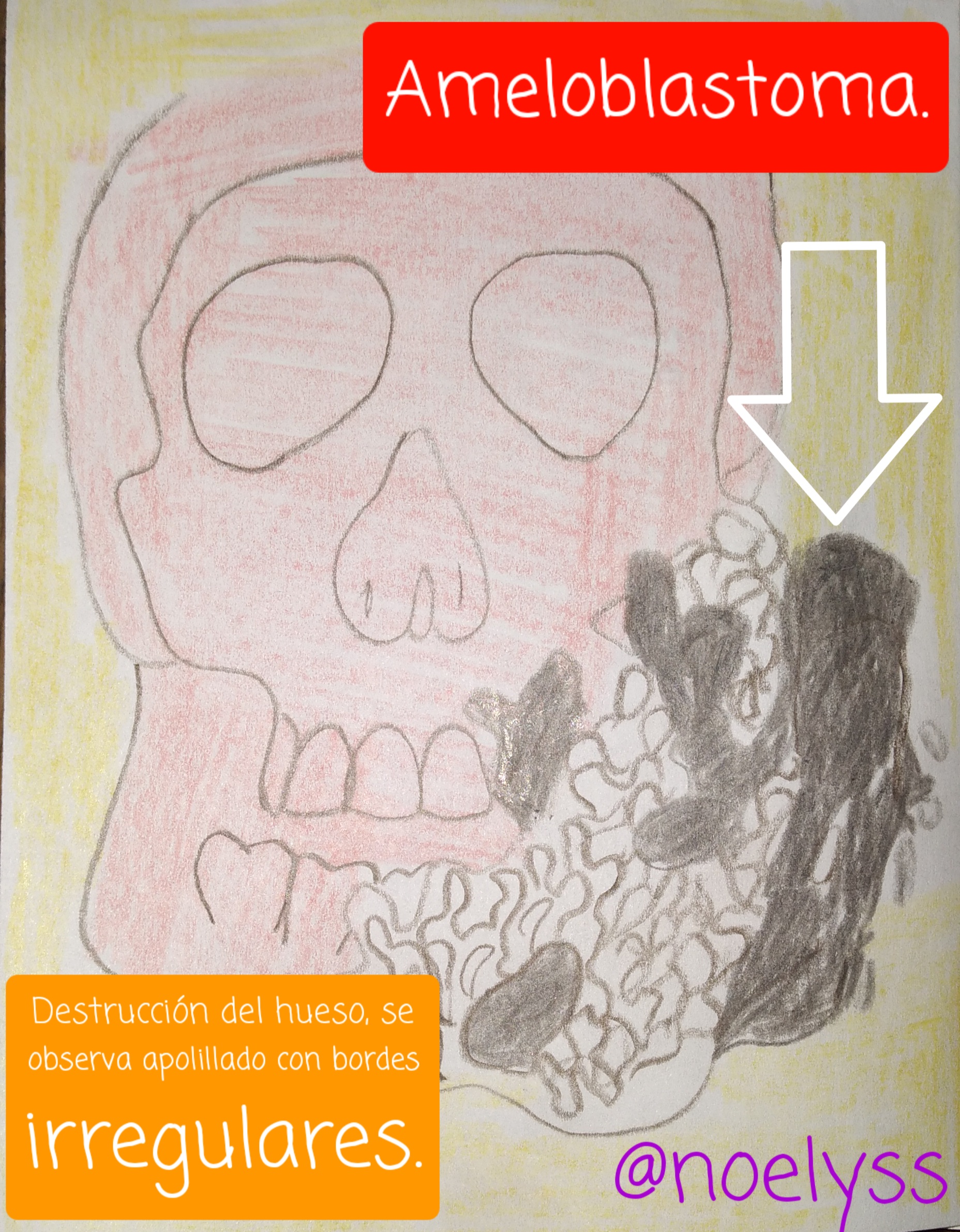
Radiograficamente se observa la destrucción ósea ya que está la va a producir la masa tumoral, verá la expansión del hueso mediante una imagen radiolúcida unilocular, si tiene un tamaño pequeño, si es grande lo veremos como una masa multilobular, su principal característica es que lo observaremos como panal de abeja o apolillado y si es de gran tamaño como burbujas de jabón.
Radiographically, bone destruction is observed as it will be produced by the tumour mass, you will see the expansion of the bone by means of a unilocular radiolucent image, if it is small in size, if it is large we will see it as a multilobular mass, its main characteristic is that we will observe it as a honeycomb or moth-eaten mass and if it is large in size it will look like soap bubbles.
Recuerda que es importante asistir periódicamente al odontólogo y tener tu radiografía panorámica, olvides que sí presentas algún bulto en tu rostro dolor o incomodidad debes asistir al odontólogo para el diagnóstico ya que lo más importante es la prevención.
Remember that it is important to visit the dentist regularly and to have your panoramic x-ray. Remember that if you have a lump on your face, pain or discomfort, you should visit the dentist for a diagnosis, as the most important thing is prevention.
---
Referencia Informativa/ Reference Informative:
✅ Esta información fue obtenida y analizada por mi gracias a la cátedra de Patología II, Universidad del Zulia Facultad de Odontología. >This information was obtained and analysed by me thanks to the chair of Pathology II, University of Zulia Faculty of Dentistry.
Los invito a formar parte de grandes comunidades como gems foodiesbeehive, stemsocial, cervantes donde podrán encontrar contenido variado desde recetas, historias, manualidades hasta increíbles post de ciencia y tecnología. Todo un universo de cultura y cripto lo encuentras aquí en Hive
 créditos @doze


Texto traducido en Deelp
Excelente post, muy organizado, original y creativo, además del tema tan importante que aborda, me encantó, felicidades.👏👏👏👏👏👏
Muchísimas gracias por tu comentario, un abrazo🤗Me gusta que te haya encantado el tema , saludos infinitos @lindoro
Mi hermana es estomatologa y la he visto de cerca estudiar mucho, lo que me ha permitido conocer técnicamente muchas cosas de esta, especialidad. Saludos y siga haciendo esos maravillosos post
Muchas gracias, saludos para tu hermana tambien🤗@lindoro
excelente información al de tu publicación amiguita, lo unico que puedo decir es que definitivamente debe ser una muy mala experiencia tener este tipo de neoplasias benignas en la cavidad oral, al final del dia debe ser muy traumático. Gracias por compartir
Muchas gracias amigo para evitar los traumas se debe asistir al especialista en cirugía bucal, en el momento preciso para tener un diagnóstico y plan de tratamiento evitando mayores consecuencias gracias por tu comentario te envío un abrazo @oswaldotorres
Wow. This was insightful. Thank you @noelyss
Always to the order to teach is the most important thing, greetings a hug thanks for commenting. @silviafx
Very nice and easy to understand. I like your illustration. Did you know Stemsocial has an art page in discord? I would love for it to work again.
From your illustration I can see that it occurs somewhere in gum.
You also wrote that it could be recurrent. How do we as doctors know a recurrent case? How do we prevent it?
I also followed you in Instagram
Hello thank you for your comment and I hope that this very well, certainly the ameloblastoma has an origin during intrauterine life, by remains of the lamina dura that are stored in the jaw, so you must take into account that sometimes it is not in us to prevent them, but certainly we can educate other people to have knowledge of this pathology and in case of observing something strange in your oral cavity or face attend an oral surgeon so they can be treated and completely removed before it is too late, greetings a hug. @ebingo
Thank you!!. Have an amazing day!!
This is very clear and straight to the point.
Can you make a diagnosis just radiographically or histology is also required?
Hola @beulah4real siempre es de vital importancia la biopsia, puesto que mediante la prueba histopatologica se corrobora la clínica, síntomas y radiografía.
Siempre es bueno conocer acerca de esos tumores y neoplasias, de como se forman y la manera de tratarlos, tan sólo para manejar dichos términos. Lo importante es poseer la confianza en quién nos trata la salud dental... Te apoyo con tu publicación y felicito tu buena disponibilidad en compartir tus conocimientos con nosotros.
El dibujo del "ameloblastoma" tiene la cara igual a "Cacique"
Saludos @noelyss