Introduction
The human arm is a complex structure that enables a vast range of movements and functions. At the heart of its versatility are the muscles, which work in concert to manipulate the environment with precision and strength. Today, we will look at the anatomy of the arm's musculature, exploring the layers of muscle that power our daily activities, from the art of writing to the demanding exertions of lifting and pushing.
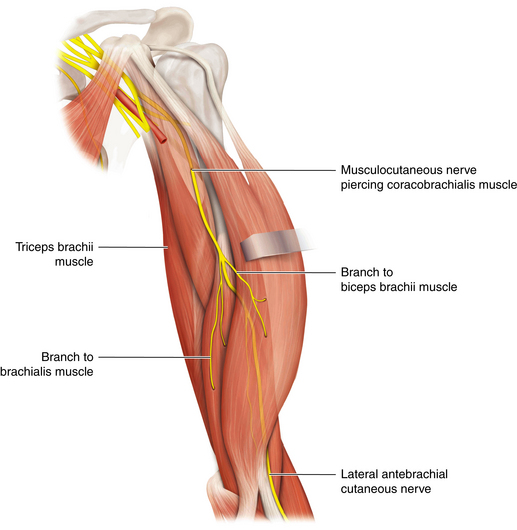
The arm's muscles are divided into two main compartments: the anterior compartment, primarily responsible for flexion and supination, and the posterior compartment, tasked with extension. These compartments house the biceps brachii, brachialis, coracobrachialis, triceps brachii, and anconeus muscles, each with unique origins, insertions, and functions. Innervated by the musculocutaneous and radial nerves, these muscles coordinate to produce the fluid and forceful movements that are needed for human function.
The Division of the Arm
The arm is anatomically divided into two main compartments: the anterior compartment and the posterior compartment. These compartments are separated by tough connective tissue known as the intermuscular septa. Imagine these septa as sturdy walls within your arm that compartmentalise and organise the muscles and nerves.
Anterior Compartment
The anterior compartment, also referred to as the flexor compartment, is primarily responsible for bending the elbow and turning the palm of the hand upward. This compartment has three significant muscles: Biceps Brachii, Brachialis, Coracobrachialis:
All these muscles are innervated by the musculocutaneous nerve, which originates from the cervical spinal nerves. The brachial artery provides the blood supply to this compartment.
Posterior Compartment
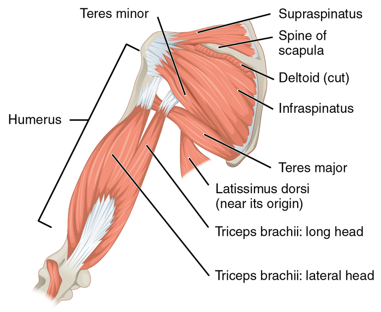
On the flip side, the posterior compartment, or the extensor compartment, enables us to straighten the elbow and bring the palm facing downward. It contains: Triceps Brachii, Anconeus
These muscles receive their nerve supply from the radial nerve and blood supply from the profunda brachii artery.
The Flexor Compartment of the Arm: A Closer Look
The flexor compartment is the anterior portion of the arm, enveloped by a sheath of connective tissue known as the brachial fascia. It's neatly partitioned from the posterior compartment by the humerus bone and the lateral and medial intermuscular septa—think of these septa as the dividers that keep everything organized within the arm.
Functional Significance:
The flexor compartment isn't just about flexing the forearm; it also contributes to the supination of the forearm—turning the palm upward—as well as aiding in the flexion of the arm at the shoulder joint. These actions are crucial for countless activities, from typing on a keyboard to performing a bicep curl at the gym.
The Biceps Brachii Muscle
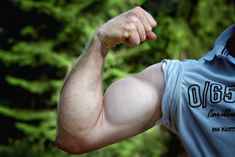
Let's turn our attention to one of the most recognized muscles of the human body—the biceps brachii. This muscle is not only an emblem of strength but also a critical component of the arm's anatomy, facilitating a range of movements.
Location and Structure:
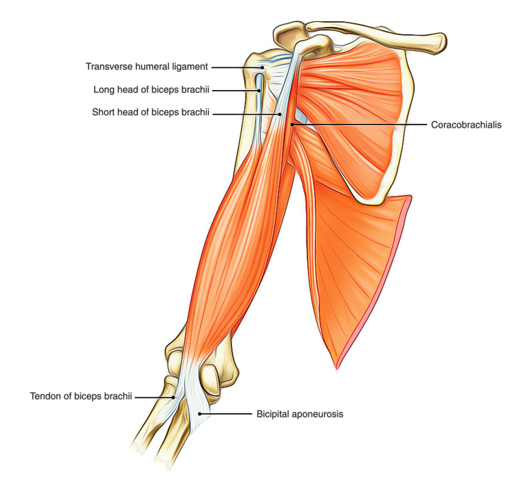
The biceps brachii is prominently situated in the front of the upper arm, between the shoulder and the elbow. It is a bipennate muscle, meaning it has two points of origin, commonly referred to as 'heads', which converge into a single tendon.
The Origin:
- Long Head: The long head originates from the supraglenoid tubercle of the scapula, travelling through the shoulder joint and down the bicipital groove of the humerus.
- Short Head: The short head arises from the apex of the coracoid process of the scapula, sharing its origin with the coracobrachialis muscle.
** Insertion**
Both heads merge to form the muscle belly, which is most visible when the muscle is contracted. This muscle belly then tapers to a single tendon that inserts into the radial tuberosity on the radius.
Innervation:
The biceps brachii is innervated by the musculocutaneous nerve, which originates from the fifth and sixth cervical spinal nerves (C5-C6). This nerve provides the electrical impulses necessary for muscle contraction.
Blood Supply:
The brachial artery, a major blood vessel of the upper arm, gives off branches that supply the biceps brachii.
Functionality:
The primary functions of the biceps brachii include:
- Flexion of the Forearm: It is the chief muscle involved in bending the elbow, bringing the forearm towards the shoulder.
- Supination of the Forearm: The biceps brachii also facilitates the rotation of the forearm, turning the palm upwards.
- Weak Flexor of the Arm: At the shoulder joint, the biceps brachii assists in the flexion of the arm, although this is not its primary role.
The Brachialis Muscle
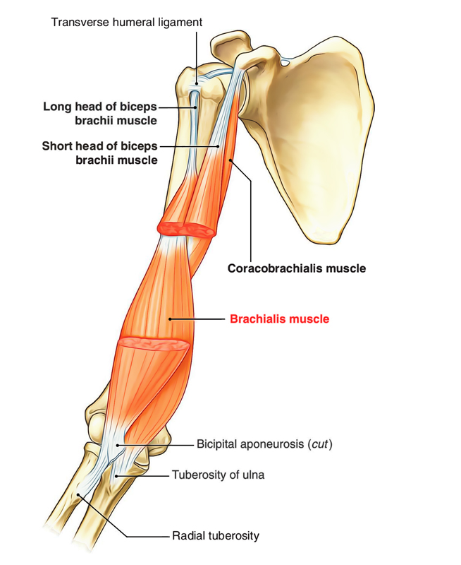
Brachialis muscle plays a crole in the flexion of our forearms. Nestled within the anterior compartment of the upper arm, the brachialis works diligently beneath the biceps brachii, contributing to the intricate machinery of our elbow joint.
Anatomy and Structure:
- Origin: The brachialis originates from the anterior surface of the distal half of the humerus. extending below to within 2.5 cm of the margin of the articular surface of the humerus at the elbow joint.
- Insertion: Its fibers converge to form a tendon, which inserts into two critical sites:
- Tuberosity of the Ulna: This bony prominence on the ulna provides a stable anchor for the brachialis.
- Anterior Surface of the Coronoid Process of the Ulna: Here, the brachialis finds another rough depression to secure its connection.
Innervation:
The musculocutaneous nerve supplies the brachialis. It runs superficially between the brachialis and the biceps brachii, ensuring the muscle receives the necessary electrical signals for contraction.
In 70-80% of people, the brachialis enjoys double innervation—courtesy of the radial nerve (C5-T1)
Blood Supply:
The brachialis receives its blood from muscular branches of the brachial artery and the recurrent radial artery.
Function:
The brachialis is the prime mover of elbow flexion. When you bend your arm, it's the brachialis that does the heavy lifting, generating about 50% more power than the biceps. Impressive, right?
Unlike the biceps, which have a hand in pronation and supination of the forearm, the brachialis remains focused on flexing the arm at the elbow joint. It's a specialist, and it takes its job seriously.
The Coracobrachialis Muscle
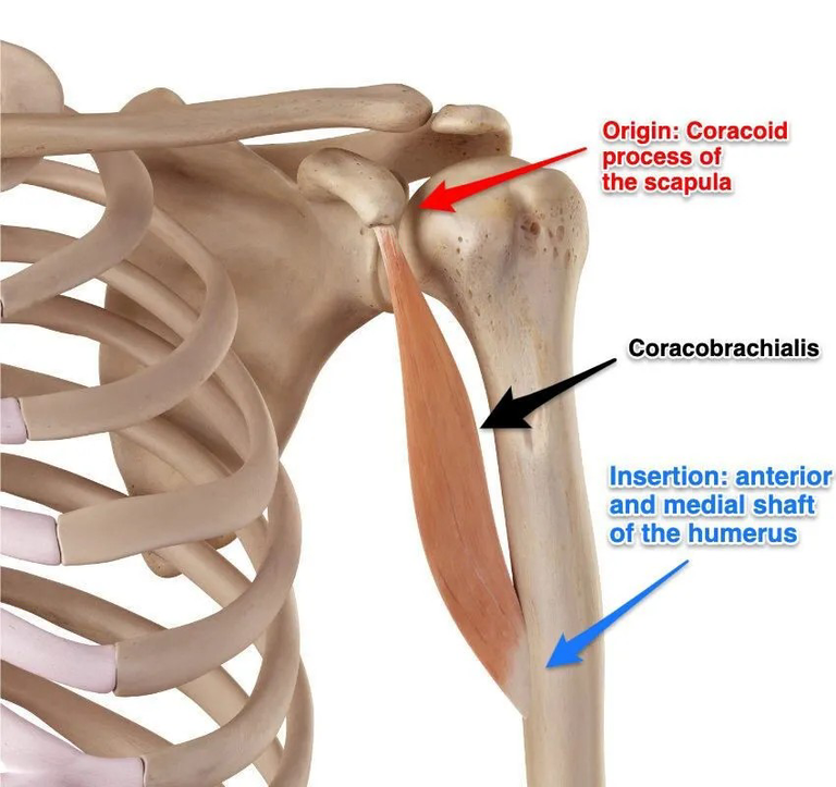
The coracobrachialis muscle,is slender yet significant within the anterior compartment of the upper arm. It operates quietly but efficiently, contributing to our arm's activities.
Anatomy and Structure:
- Origin: The coracobrachialis originates from the deep surface of the coracoid process of the scapula—that small, hook-like structure near the shoulder joint.
- Insertion: From its origin, the muscle fibers extend inferolaterally, attaching to the anteromedial surface of the humeral shaft.
Relations:
The coracobrachialis lies posterior to the pectoralis major muscle and anterior to the tendons of subscapularis, latissimus dorsi, teres major, and the medial head of triceps. It connects these structures, ensuring smooth communication between them.
Medially, it cozies up to the biceps brachii and brachialis muscles. And guess what? The median nerve crosses right in front of its humeral insertion.
If you want to feel the coracobrachialis, palpate the lateral border of the axilla—it's there, waiting to be discovered.
Innervation:
The musculocutaneous nerve (C5-C7), supply the muscle
Blood Supply:
The brachial artery supply blood to the coracobrachialis. Additional support comes from the anterior circumflex humeral and thoracoacromial arteries.
Function:
When it comes to action, the coracobrachialis has two main moves:
Flexion of the Arm: Imagine lifting a heavy bag or waving hello—this muscle is behind those movements.
Adduction of the Arm: It pulls the arm toward the trunk, like a friend urging you to join the dance floor or actions like that.
The coracobrachialis supports the biceps brachii and brachialis.
Posterior Compartment of the Arm
The posterior compartment is one of the two main compartments in the arm, separated from the anterior compartment by the humerus and the intermuscular septa.
This compartment is home to the muscles responsible for the powerful extension movements of the arm: Triceps Brachii, Anconeus
Innervation and Blood Supply:
Innervation: The radial nerve (C7, C8) is the primary nerve that innervates the muscles of the posterior compartment, providing the necessary signals for muscle contraction
Blood Supply: The profunda brachii artery, along with the superior and inferior ulnar collateral arteries, ensures a rich blood supply to these muscles
Functional Importance:
The posterior compartment plays a critical role in extending the forearm, enabling us to perform actions like pushing open a door or performing a push-up. The coordinated action of the triceps brachii and anconeus allows for these movements to be executed with strength and precision.
As we proceed, we will explore each muscle within this compartment in detail, understanding their specific anatomical features, functions, and clinical significance.
Triceps Brachii Muscle
Triceps brachii muscle is the sole constituent of the posterior compartment of the arm and functions in arm extension. This muscle is unique in its structure and function.
Anatomical Structure:
The triceps brachii is a large, three-headed muscle that spans almost the entire length of the humerus. Each head has its own origin:
- Long Head: Originates from the infraglenoid tubercle of the scapula. It extends slightly above to the adjacent glenoid labrum and blends with the glenohumeral capsule, contributing to the shoulder joint's stability.
- Lateral Head: Arises from a narrow ridge on the posterior surface of the humerus, just superior to the radial groove. Some fibers also arise from the lateral intermuscular septum¹.
- Medial Head: Overlapped by the long and lateral heads, it has a broad origin along the entire posterior surface of the humerus inferior to the radial groove.
These heads converge into a common tendon that inserts into the olecranon of the ulna.
Innervation:
The triceps brachii is innervated by the radial nerve (C6-C8), which provides the necessary signals for muscle contraction
Blood Supply:
The muscle receives oxygen and nutrients from the deep brachial artery and the superior ulnar collateral artery
Function:
The triceps brachii has several key functions:
- Elbow Joint: It is the principal muscle responsible for the extension of the forearm at the elbow joint.
- Shoulder Joint: The long head also contributes to the extension and adduction of the arm at the shoulder joint.
Anconeus Muscle
Origin: The anconeus muscle originates from the dorsal aspect of the lateral epicondyle of the humerus, just proximal to the common extensor tendon. Its tendon lies deep to the muscle belly of the extensor carpi radialis longus and is partially attached to the dorsal capsule of the humeroulnar joint.
Insertion: The anconeus tendon spreads out obliquely and medially into a wide muscle belly, inserting at the lateral surface of the olecranon of the ulna and the adjoining posterior surface of the ulnar shaft.
Structure: Despite its small size, the anconeus has a flat, somewhat fan-shaped structure, with fibers running obliquely from its origin to its insertion points.
Innervation: The anconeus muscle receives its innervation from a motor branch of the radial nerve (C7-C8).
Blood Supply: It is supplied by the recurrent interosseous branch of the posterior interosseous artery, along with contributions from a small number of musculocutaneous perforators.
Function:
- Assists in Forearm Extension at the Elbow Joint:
- The anconeus contributes to the extension of the forearm at the elbow joint. When you straighten your arm, it quietly supports the action alongside the triceps brachii.
- Stabilization of the Elbow Joint:
- Beyond extension, the anconeus provides support for both the dorsal capsule of the humeroulnar joint and the ulna itself. It helps maintain stability during various movements.
Please, do not forget to consult your Atlas textbooks where necessary
This is the end of this topic. See you again next week!!
References
(1) Anconeus muscle: Origin, insertion, innervation, function | Kenhub. https://www.kenhub.com/en/library/anatomy/anconeus-muscle.
(2) Anconeus - Attachments - Actions - TeachMeAnatomy. https://teachmeanatomy.info/encyclopaedia/a/anconeus/.
(3) Anconeus Muscle Origin, Anatomy & Function | Body Maps - Healthline. https://www.healthline.com/human-body-maps/anconeus-muscle/male.
(4) Triceps brachii muscle: Attachments, supply and functions - Kenhub. https://www.kenhub.com/en/library/anatomy/triceps-brachii-muscle.
(6) Triceps brachii - Physiopedia. https://www.physio-pedia.com/Triceps_brachii.
(7) Anatomy of posterior compartment of the arm | PPT - SlideShare. https://www.slideshare.net/DrMohammadMahmoud/anatomy-of-posterior-compartment-of-the-arm.
(8) Muscle compartments of the upper arm | Complete Anatomy - 3D4Medical. https://3d4medical.com/blog/muscle-compartments-of-the-arm.
(9) Coracobrachialis: Attachments, innervation, function. | Kenhub. https://www.kenhub.com/en/library/anatomy/coracobrachialis-muscle.
(10) Coracobrachialis - Attachments - Actions - TeachMeAnatomy. https://teachmeanatomy.info/encyclopaedia/c/coracobrachialis/.
(11) Coracobrachialis muscle - Wikipedia. https://en.wikipedia.org/wiki/Coracobrachialis_muscle.
(12) Brachialis muscle - Wikipedia. https://en.wikipedia.org/wiki/Brachialis_muscle.
(13) Brachialis muscle: Location, origin and insertion, action | Kenhub. https://www.kenhub.com/en/library/anatomy/brachialis-muscle.
(14) Biceps brachii muscle: Origin, insertion, action | Kenhub. https://www.kenhub.com/en/library/anatomy/biceps-brachii-muscle.
(15) Biceps Brachii - Attachments - Actions - TeachMeAnatomy. https://teachmeanatomy.info/encyclopaedia/b/biceps-brachii/.
(16) Biceps brachii - Location, Structure, Diagram, Function. https://anatomy.co.uk/biceps-brachii/.
(17) Flexor compartment of arm - Medical Dictionary. https://medical-dictionary.thefreedictionary.com/flexor+compartment+of+arm.
(18) Arm muscles: Anatomy, attachments, innervation, function | Kenhub. https://www.kenhub.com/en/library/anatomy/arm-muscles.
(18) Muscles of the Anterior Forearm - Flexion - TeachMeAnatomy. https://teachmeanatomy.info/upper-limb/muscles/anterior-forearm/.
(19) Fascial compartments of arm - Wikipedia. https://en.wikipedia.org/wiki/Fascial_compartments_of_arm.
(20) Arm muscles: Anatomy, attachments, innervation, function | Kenhub. https://www.kenhub.com/en/library/anatomy/arm-muscles.
(21) Muscles of Arm (Left) | Complete Anatomy - Elsevier. https://www.elsevier.com/resources/anatomy/muscular-system/muscles-of-upper-limb-left/muscles-of-arm-left/21670.
(22) Arm muscles: Anatomy, attachments, innervation, function | Kenhub. https://www.kenhub.com/en/library/anatomy/arm-muscles.
(23) Muscles of the Upper Arm - Biceps - Triceps - TeachMeAnatomy. https://teachmeanatomy.info/upper-limb/muscles/upper-arm/.
(24) Anatomy of the arm: Video, Anatomy & Definition | Osmosis. https://www.osmosis.org/learn/Anatomy_of_the_arm.
(4) Video: Muscles of the arm | Kenhub. https://www.kenhub.com/en/videos/muscles-of-the-arm.


I am a complete beginner who resides in Africa's Western Hemisphere. My name is James, but you may reach out to me through the Facebook page [James Kossy] (https://www.facebook.com/christ.messenger.904) Physics, chemistry, and biology are the three topics that I find most enjoyable. My current studies are taking place at the university level, with the intention of becoming a recognized professional in physiotherapy. I am fascinated by all things technological, and I take pleasure in contributing to the fascinating technological advancements that are taking place throughout the world today. In my spare time, I'd like to learn more about programming and help others with any technical problems they may be having. 💞 ***🌹❤️ Thank you so much to everyone who has supported me thus far. ****💞 At the moment, I don't have the right words to say how much I appreciate all of your help. You never cease to astonish me with your generosity. For me, this has turned into a haven of enjoyment. Thanks to colleagues like you, this has all been possible. You've been a great support for me. Everything you have done for me and my family has been greatly appreciated, and I will always be grateful to you. 💕.
Posted Using InLeo Alpha
Thanks for your contribution to the STEMsocial community. Feel free to join us on discord to get to know the rest of us!
Please consider delegating to the @stemsocial account (85% of the curation rewards are returned).
You may also include @stemsocial as a beneficiary of the rewards of this post to get a stronger support.