The kidneys are the organs of the human body that perform many functions for homeostasis, which is primarily as an excretory organ and a balance regulator of fluids and acid-base in the body. There are a pair of kidneys in humans, each on the left and right (lateral) vertebral bones and located retroperitoneal (behind the peritoneum). In addition, a pair of kidneys is also equipped with a pair of ureter, a vesika urinaria (bladder / bladder) and urethra that carries urine to the environment outside the body.
Kidney
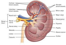
The kidney is a nut-shaped organ, there is a pair (each one on the right and left of the vertebra) and its position is retroperitoneal. The right kidney lies slightly lower (approximately 1 cm) than the left kidney, this is due to the liver that pushes the right kidney. The left upper renal pole is the upper edge of the rib 11 (T12 vertebra), whereas the right upper renal pole is the lower edge of rib 11 or rib 12. The left renal pole is the transverse process of the L2 vertebra (approximately 5 cm from the iliac crest) and the polar Under the right kidney is the mid-L3 vertebra. From these limits it can be seen that the right kidney is inferior to the left kidney.
In general, the kidney consists of several parts:
The cortex, the portion of the kidney where it contains / consists of the renal / Malpighi corpus (glomerulus and Bowman capsule), proximal con- tractal tubules and distal contrast tubules
Medulla, which consists of 9-14 pyiramid. It consists of the rectus tubule, the loop of Henle and the collecting tube (ductus colligent).
Columna renalis, the cortical part between the renal pyramid
The renal process, the pyramid / medulla portion, protrudes toward the cortex
Hilus renalis, which is a part / area where blood vessels, nerve fibers or ducts enter / leave the kidney.
Papilla renalis, the connecting part of the collecting duct and the calix minor.
Calix minor, the branching of calix major.
Calix major, the branching of the renal pelvis.
The renal pelvis, also called the kidney cup, is the part that connects the calix major and the ureter.
Ureter, the channel that carries urine to vesica urinaria.
Urine Formation Process
In a previous post titled Excretion System In Humans, we have discussed little about the kidneys and parts of it. As we know, the kidney is one of the excretory organs that function to remove metabolic waste substances in the form of urine. The process of urinating itself is divided into three stages, namely filtration stage, reabsorption stage, and augmentation stage. And here is the explanation.
Terjemahan
Matikan terjemahan instan
3185/5000
- Filtering (Filtration)
Firstly, water containing water (H2O), glucose (C6H12O6), ammonia (NH3), salt, urea, and amino acids enter the glomerulus through the afferent arteriole to undergo a filtration process. Glomerulus is part of the malpighi body. These glomerular capillary cells that possess porous and high-pressure characteristics further facilitate the filtration or filtration process.
In the glomerulus, the process of re-absorption of blood cells, blood chips, and protein molecules are large. Meanwhile, small molecules contained in the blood such as glucose, amino acids, sodium, potassium, chloride, bicarbonate, and urea pass from filtering and coalesce along with the primary urine. The primary urine that has been formed will then be accommodated inside the bowman capsule.
- Reabsorption (Reabsorption)
After the blood undergoes filtration in the glomerulus, the primary urine that has been accommodated in the bowman capsule will enter into the proximal contralus tubule for reabsorption.
The primary urine formed through the filtration process still contains some substances that are useful to the body, such as glucose, amino acids, and some ions such as Na +, Cl-, HCO3-, and K +. Substances that are still useful for this body will then go into the blood vessels that surround the tubules. Among the substances that are no longer useful to the body such as ammonia, salt, and urea will form secondary urine. This secondary urine then enters the henle arch to get to the distal contrast tubule. As it passes through the henle curve, the urine becomes more concentrated and the volume decreases due to terosmosis. In this secondary urine, is no longer found substances that are still useful for the body. Meanwhile, the composition of metabolic waste substances will increase.
- Augmentation
After undergoing reabsorption, the secondary urine will enter the distal contrast tubule through the henle arch. In the distal contrast tubule, the secondary urine will lose a lot of water (H2O) so the urine becomes more concentrated. Here also the secondary urine has the addition of residual substances and toxic substances such as hydrogen ions (H +) and urea.
After experiencing the addition of various waste substances in the augmentation process, the secondary urine then to the pelvis and then into the vesica urinaria through the ureter channel to be temporarily accommodated. From there urine will go to the bladder. The bladder is only able to accommodate approximately 300 ml of urine. When the bladder is fully charged, then the wall of the bladder will be depressed so we feel the urge to urinate.
Urine is stored in the bladder will then exit the body through the urethral tract. This real urine has a 96% composition of water, 2.5% urea, 1.5% salt, and has also been mixed with bile dyes that give color to urine.
Man himself will normally produce urine as much as two liters per day. Many of the least urine produced by humans actually also caused by several factors such as the amount of water drunk, air temperature, and blood pressure.