This is a case of a 66 year old male managed as community acquired pneumonia with pleural effusion. Thoracentesis was done and the pleural fluid was sent for cytologic examination. I took these pictures using my phone's cam through the new microscope where instead of an objective lens we have an LCD monitor. The company wanted use to use the microscope as a demo.
These are poor quality images as the lighting conditions when I took it were night time and our office's bulbs need replacement. It's that or I'm just bad at taking pictures.
I'm going to give you the spoiler here that the pictures show malignant cells. No lung mass was identified on imaging and it's a common scenario where people come it for benign conditions like chronic cough only to get an incidental finding of malignancy.
Here's the show and tell part.
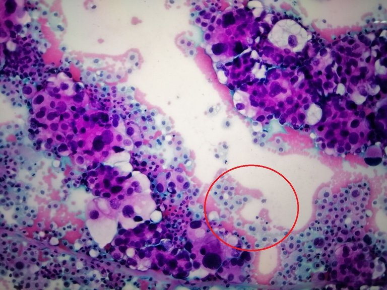
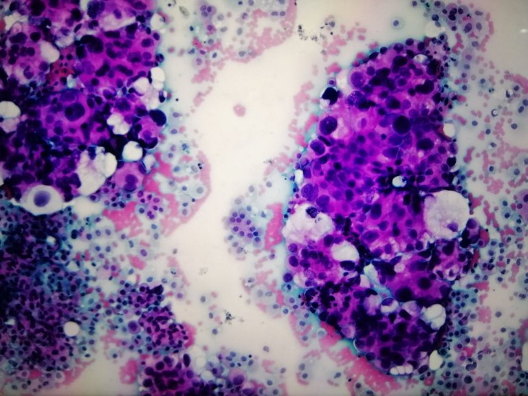
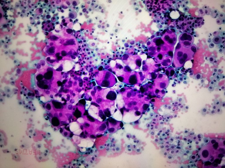
Taken at low power view, I highlighted in red circle the normal mesothelial cells lining the pleural epithelium. The elephant in the room are those large irregular cells in clusters surrounding the red blood cells and mesothelial cells. This is just for comparison. I don't get these type of smears often but when I do, it makes the diagnosis quicker knowing the answer already stares back at you.
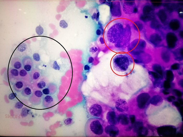
Normal cells in black while the malignant cells in red circles showing mitosis (metaphase and telophase). We don't really need to see mitotic figures to confirm malignancy. Mitosis means the cells are actively dividing and given that the cells dividing are pleomorphic, it just adds proof that we're dealing with malignant cells.
The description I gave here are tumor cells having hyperchromatic, markedly pleomorphic nuclei with inconspicuous to prominent nucleoli, fine to coarse chromatin pattern with moderate to ample cytoplasm.
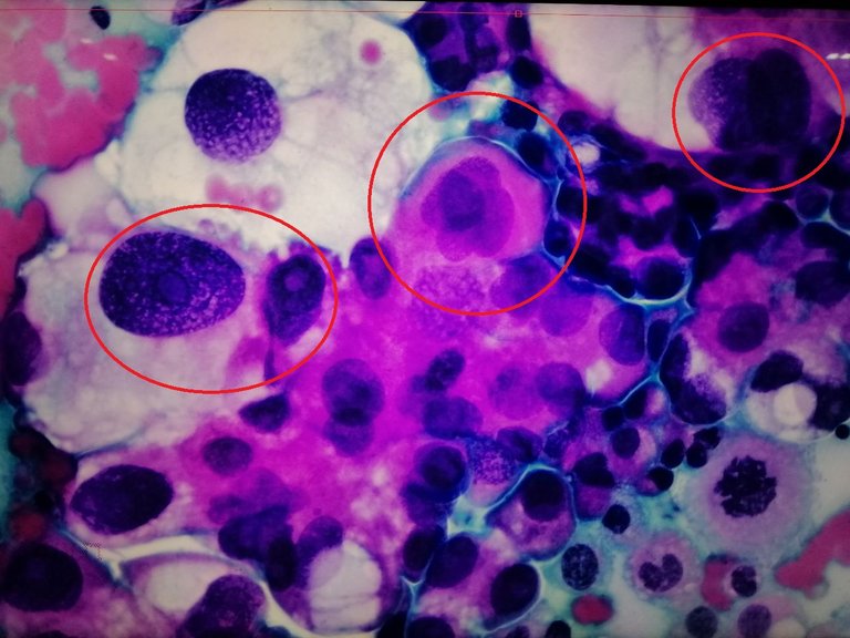
The center red circle shows a malignant cell with multiple nuclei. The left one show a prominent nucleoli (mean eye). The darkened stains on their nuclear membrane can be attributed to how much cellular activity is going on compared to the lighter stained cells which aren't mitotically active. I missed the lower right corner cell where it looks like an irregular raisin nuclei but that's probably a mitotic figure or an apoptotic cell.
The patient has no lung mass on imaging but that doesn't mean there isn't any tumor on the lungs. This could be from somewhere else or within the lungs but too small to be recognized.
We don't immediately say it's a lung cancer because the only thing we are sure of is that there are malignant cells in the pleural fluid. It could still be from somewhere else. That's why immunohistochemical studies were requested to determine the primary site.
If it is indeed from the lungs, the prognosis is usually bad, well any cancer is generally bad but lung cancers tend to be more aggressive.
If you made it this far reading, thank you for your time.
Posted with STEMGeeks
That sucks for the patient.
I wasn't expecting the malignant cells to be so clear and distinct on a slide.
!discovery 41
It makes the job easier when these things show up at low power but it definitely sucks for the patient who only thought they had tuberculosis or pneumonia.
Very descriptive and interesting information. Very good post.
Congratulations @adamada.stem! You have completed the following achievement on the Hive blockchain And have been rewarded with New badge(s)
Your next target is to reach 90 posts.
You can view your badges on your board and compare yourself to others in the Ranking
If you no longer want to receive notifications, reply to this comment with the word
STOPTo support your work, I also upvoted your post!
Check out our last posts:
This post was shared and voted inside the discord by the curators team of discovery-it
Join our Community and follow our Curation Trail
Discovery-it is also a Witness, vote for us here
Delegate to us for passive income. Check our 80% fee-back Program