Greetings esteemed colleagues, Welcome to another significant installment of the study of human anatomy. The focus of my discussion today pertains to the thoracic wall and the contents that are situated within this anatomical region.
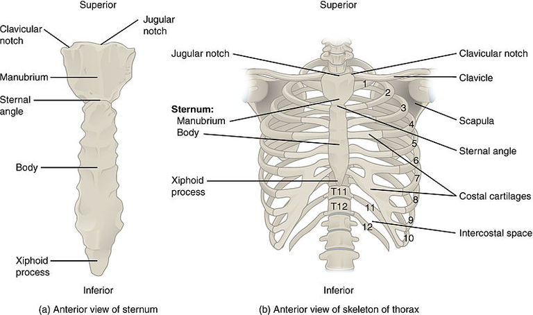
Twelve thoracic vertebrae, which laterally give birth to ribs that surround the lateral and anterior thoracic cavities, form the bony structure of the thoracic wall. The manubrium and sternum are connected by the first nine ribs, which arc around the lateral thoracic wall. The costal margins of the ribcage just above them are where ribs 10 and 12 connect. Because of their short paths, ribs 10 and 12 do not extend to the chest.
The manubrium and the body of the sternum are immediately connected by the first seven ribs, which are referred to as "real ribs." Only the eighth through tenth ribs use the costal cartilages to connect to the lower portion of the sternum. The 11th and 12th ribs are known as "floating ribs" because they are not physically connected to the ribcage. Because they don't directly connect to the ribcage, ribs eight through ten are referred to as "artificial ribs." The costal facet of two opposite vertebrae and the ribcage articulate at the level of the spine. Each rib's head is encased in an articular capsule, and the radiate ligament helps to form a connection to the transverse process. The ribs slightly curve around the lateral thoracic wall once they have separated from the vertebrae and are moving toward the anterior wall of the thoracic chamber.
The anterior chest wall is defined by the sternum, the straight breast bone. The thick manubrium, the lengthy body of the sternum, and the xiphoid process are the three distinct bone parts that make up the sternum, each of which is distinctive in size and form. It grows separately from the ribcage. The ribcage may occasionally not develop completely, exposing the heart beneath.
The manubrium, which is the most prominent part of the ribcage, is also the first part of the embryo to develop. The manubrium develops quickly afterwards, followed by the sternal body and xiphoid process. The manubrium is anatomically situated between the thoracic spinal segments T3 and T4. The manubrium is part of the ribcage that is both the broadest and thickest. The suprasternal groove is one aspect of the manubrium that stands out during a physical examination of the breast. One can sense the strong clavicle connection on both sides of this notch. A midline cut in the manubrium is all that some lung surgeons will use to gain entry to the upper mediastinum, suprasternal goitre, or thymus.
The region of the spinal bodies T5–T9 is where the sternal body is situated. It is very robust and encompasses a good deal of the mid-chest. Using a mechanical saw, doctors typically cut through the sternum to gain entry to the thoracic region.
The xiphoid process is a delicate bone that is very tiny. Its dimensions can range from two to five centimetres, and its form can change. The shape of the xiphoid can be circular, bifid, or curled inwards or outwards. The xiphoid is mostly cartilaginous when people are younger, but by the time they are 40, it has nearly completely ossified. The xiphoid is almost definitely fully calcified by the time a person is 60 years old or older. The heart is only a few fingerbreadths below the xiphoid, so to conduct pericardiocentesis securely, the needle must be put immediately there.
Arrangement and Purpose
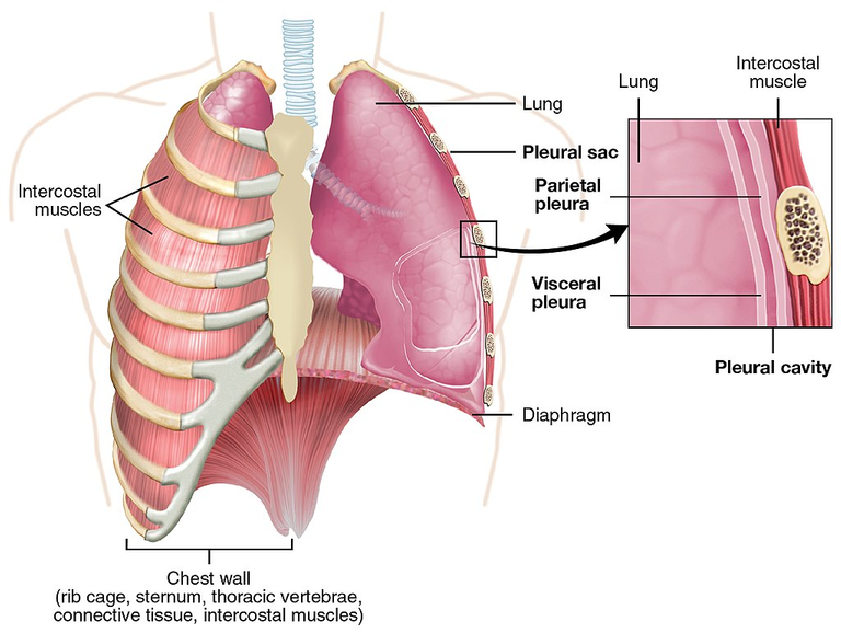
The mediastinum and two pleural chambers, one on either side, make up the three sections that make up the thoracic cavity. The lungs are located in the pleural spaces, while the heart and great arteries are located in the mediastinum. The muscles of the thorax, upper limbs, back, and belly are attached to the thoracic cage, which also safeguards the heart and lungs. It connects to the neck superiorly via the thoracic exit and is divided from the belly inferiorly by the respiratory diaphragm.
Clinicians and doctors use the limits of the thoracic wall as crucial markers for a variety of operations, such as sternotomy, pericardiocentesis in patients with cardiac tamponade, and thoracentesis for pleural effusion. The sternum and costal cartilages in the front, the ribs and intercostal gaps in the middle, the thoracic vertebrae and intervertebral discs in the back, the suprapleural membrane in the top, and the breathing diaphragm in the bottom, define the boundaries of the thoracic wall.
Development
As the paraxial mesoderm begins to spiral into an orderly cell known as a somitomere, somite formation commences. To create individual somites, these somitomeres detach from the presomitic paraxial mesoderm and group together by epithelium. The division of the somite results in the formation of the skin's dermis as well as the cartilage of the vertebrae, ribcage, and rib cage muscle.
The flow of Blood and Lymphatics
Each intercostal area is supplied by two branches of the anterior intercostal vessels and the posterior intercostal artery. Between the interior and deepest intercostal muscles in the costal groove, these intercostal blood vessels travel alongside the nerves. The vein, artery, and nerve are organized from superior to lower.
The superior (supreme) intercostal artery supplies blood to the posterior intercostal artery for the first two intercostal areas. The costocervical stem of the subclavian artery is where this vessel originates. A pair of subcostal arteries and the final pair of posterior intercostal vessels from the third through the eleventh intercostal spaces arise straight from the descending thoracic aorta.
The internal thoracic artery, which arises from the first segment of the subclavian artery, gives rise to the anterior intercostal vessels of the first through sixth intercostal areas. The musculophrenic artery, a final tributary of the internal thoracic artery, bears branches in the 7th through 9th intercostal regions. In the costal cleft, the anterior and posterior intercostal arteries anastomose horizontally.
The corresponding anterior intercostal veins empty into internal thoracic or musculophrenic veins, while the corresponding posterior intercostal veins drain into azygos or hemiazygos veins. The parasternal and intercostal lymph nodes receive drainage from the chest wall's lymphatics. While the intercostal nodes from the lower thorax drain into the thoracic duct, the parasternal lymph nodes and intercostal lymph nodes from the upper thorax empty into the bronchomediastinal trunk.
Muscles

There are three intercostal muscles that work together to help with breathing: the externally intercostal, internal intercostal, and innermost intercostal muscles. These muscles are located in the intercostal spaces and how the intercostal nerves and blood vessels run between them. The external intercostal muscle is the first layer. It's great to know that the external intercostal muscles play an important role in breathing by extending from the rib tubercle to the costochondral junction. The anterior intercostal membrane takes over where the muscle fibers end, ensuring a smooth transition.
The internal intercostal muscle is an important part of the intermediate layer. This muscle is amazing! It extends all the way from the sternum to the rib cage, and its fibers are replaced by the posterior (internal) intercostal membrane. It's amazing how the innermost intercostal muscle forms the deepest layer and is lined internally by the endothoracic fascia, which is then lined internally by the parietal pleura.
Nerves supply
The intercostal nerves, which are the anterior rami of spinal nerves T1-T11 and the anterior ramus of T12, innervate the thoracic wall mainly. A dermatome and a myotome are supplied by each intercostal nerve. The remaining intercostals do not make a plexus; only the anterior ramus of T1 forms the lower stem of the brachial plexus.
The exterior intercostal, internal intercostal, and deepest intercostal muscles are the three intercostal muscles. The intercostal nerves and blood arteries connect these muscles with the intercostal regions where they are located. The external intercostal muscle is the tissue that is the most basic. The anterior (external) intercostal membrane replaces the muscle fibres at the costochondral joint where the external intercostal muscles stretch posteriorly from the rib tubercle.
The middle stratum is created by the interior intercostal muscle. This muscle runs posteriorly from the sternum to the rib cage, where the posterior (interior) intercostal membrane replaces the muscle fibres. The endo thoracic fascia, which in turn is bordered inwardly by the parietal pleura, lines the innermost intercostal muscle, which makes up the deepest stratum.
Clinical Relevance
The thoracic region is predisposed to being a location of high clinical importance due to the liveliness of the organs, vessels, and nerves there.
Due to its lifelong presence of hematopoietic marrow, the sternum is frequently used as a location for bone marrow extraction. The left brachiocephalic vein in the higher portion and the aortic arch in the lower part of the manubrium are two structures connected to the rear surface that can be punctured if the sternal puncture is performed incorrectly.
The physician will conduct a diagnostic treatment called thoracentesis to drain extra fluid from the thoracic chamber for either diagnostic or therapeutic reasons. Depending on the patient's comfort, this operation can be carried out in one of two postures. When the patient is upright or seated, the needle is typically placed posteriorly between the 9th and 10th ribs in the mid-scapular line, while when the patient is lying supine, the needle is typically placed in the midaxillary line between the 6th and 8th ribs. Nonetheless, in either scenario, inadequate needle insertion may lead to the perforation of the liver or spleen.
References
- Thoracic wall - Wikipedia. (2021, November 1). Thoracic Wall - Wikipedia. https://en.wikipedia.org/wiki/Thoracic_wall
- Thorax. (n.d.). Thorax: Anatomy, Wall, Cavity, Organs & Neurovasculature | Kenhub. https://www.kenhub.com/en/library/anatomy/thorax
- Hussain, A., & Burns, B. (2022, July 25). Anatomy, Thorax, Wall - StatPearls - NCBI Bookshelf. Anatomy, Thorax, Wall - StatPearls - NCBI Bookshelf. https://www.ncbi.nlm.nih.gov/books/NBK535414/#:~:text=The%20thoracic%20wall%20consists%20of,to%20the%20manubrium%20and%20sternum.
- Thoracic Muscles - Attachments - Actions - TeachMeAnatomy. (n.d.). Thoracic Muscles - Attachments - Actions - TeachMeAnatomy. https://teachmeanatomy.info/thorax/muscles/thoracic-cage/
- Thoracic cavity | Description, Anatomy, & Physiology. (n.d.). Encyclopedia Britannica. https://www.britannica.com/science/thoracic-cavity
- Chest Wall: Anatomy | Concise Medical Knowledge. (2023, March 29). Lecturio. https://www.lecturio.com/concepts/chest-wall/
- Cook, M. S., & Weinhaus, A. J. (n.d.). Anatomy of the Thoracic Wall, Pulmonary Cavities, and Mediastinum. Anatomy of the Thoracic Wall, Pulmonary Cavities, and Mediastinum | SpringerLink. https://doi.org/10.1007/978-3-319-19464-6_4
- Chapter 2. Anterior Thoracic Wall | The Big Picture: Gross Anatomy | AccessPharmacy | McGraw Hill Medical. (n.d.). Chapter 2. Anterior Thoracic Wall | the Big Picture: Gross Anatomy | AccessPharmacy | McGraw Hill Medical. https://accesspharmacy.mhmedical.com/content.aspx?bookid=381§ionid=40140008
- Samy, D. I. (2001, March 1). Thoracic wall. Thoracic Wall. https://www.slideshare.net/IhabSamy/thoracic-wall


Thanks for your contribution to the STEMsocial community. Feel free to join us on discord to get to know the rest of us!
Please consider delegating to the @stemsocial account (85% of the curation rewards are returned).
You may also include @stemsocial as a beneficiary of the rewards of this post to get a stronger support.
My dear bro James!
Really great medical information!
By the way, can you take a human anatomy class in the future?
It is common for many medical students to quit becoming doctors because they cannot stand the pain of human anatomy classes.
Well, anatomy required a long time of concentration and constant study for you to scale through in the course.
Lol, we had an anatomy practical today and we dissected cadavers
Dear my young bro James!
you are a very strong person I won't touch a human corpse. Perhaps, I'll have nightmares every night from human corpse!😨
Congratulations your publication has been chosen among the best of the day.
KEEP CREATING GOOD CONTENT.
Thank you so much...
Informative, interesting and, above all, educational. Seeing how you explain the human anatomy here at Hive is simply great. What a great post you have done!
Thank you so much for stopping by. i'm glad you found the article really interesting
Absolutely, I mean. It's health related and has well writing also. Thank you for made this, @jsalvage