Introduction
Have you ever wondered how your arms and hands work? How do you lift, bend, twist, and grip things with such ease and precision? The answer lies in the structure and function of your upper limbs. In this article, we will explore the basics of upper limb anatomy, and why it is important to understand it.
Upper limb anatomy refers to the study of the bones, muscles, nerves, blood vessels, and other tissues that make up your arms, from your shoulders to your fingertips. It is a topic that has many applications in medicine and education. For instance, upper limb anatomy can help you:
- Learn how to prevent and treat injuries and diseases that affect your arms and hands, such as fractures, arthritis, carpal tunnel syndrome, and more.
- Appreciate the diversity and beauty of human anatomy, and how it reflects our evolution, adaptation, and variation.
- Enhance your skills and knowledge in various fields and disciplines, such as art, sports, engineering, robotics, and more.
By the end of this article, you will have a better understanding of the main components and functions of your upper limbs, and how they work together to enable you to perform various tasks and activities. You will also learn some fun and interesting facts about upper limb anatomy that you may not have known before.
Before we get started, let me inform you that this topic will definitely linger to a great extent. Hence we'll be taking the topics under the upper limb bit by bit so you don't get overwhelmed.
So let's get started!
Overview of the upper limb

The upper limb is an important part of the human body, allowing us to perform a wide range of motions and activities. Consisting of the shoulder, arm, elbow, forearm, wrist, and hand, the anatomy of the upper limb is a complex and intricate system of bones, muscles, tendons, ligaments, and nerves that work together to provide strength, flexibility, and dexterity.
At the top of the upper limb is the shoulder, which is made up of three bones: the clavicle (collarbone), scapula (shoulder blade), and humerus (upper arm bone). The shoulder joint is a ball-and-socket joint that allows for a wide range of motion, including flexion, extension, abduction, adduction, and rotation. The muscles and tendons surrounding the shoulder joint provide stability and support, enabling us to lift, push, pull, and rotate our arms.
The arm consists of the humerus, the largest bone in the upper limb, and is responsible for bending and straightening the elbow joint. The biceps and triceps muscles are the primary movers of the arm, allowing for flexion and extension of the elbow. The elbow joint is a hinge joint that provides stability and support for activities such as lifting, carrying, and throwing.
Moving down the arm, we come to the forearm, which is made up of two bones: the radius and ulna. These bones are responsible for rotating the wrist and hand, as well as flexing and extending the fingers. The muscles in the forearm, such as the flexor and extensor muscles, allow for precise and coordinated movements of the wrist and fingers, essential for activities such as writing, typing, and playing musical instruments.
The wrist and hand are structures, consisting of multiple small bones, ligaments, tendons, and nerves. The wrist joint allows for flexion, extension, abduction, and adduction of the hand, while the hand itself has intricate movements such as grasping, gripping, and fine motor skills. The muscles and tendons in the hand and wrist work in harmony to provide strength and precision, enabling us to perform tasks such as holding a pen, tying shoelaces, and using tools.
In addition to the musculoskeletal structures, the upper limb also contains an extensive network of nerves that provide sensation and control to the entire arm and hand. The brachial plexus is a network of nerves that originates from the spinal cord and innervates the upper limb, allowing for voluntary and involuntary movements, as well as sensory perception.
In conclusion, the anatomy of the upper limb is a system that allows us to perform a wide range of activities and tasks. Its arrangement of bones, muscles, tendons, ligaments, and nerves provides strength, flexibility, and dexterity, crucial for daily functioning and quality of life.
The Clavicle: The key Bone for the Upper Limb
To begin our journey on the upper limb, let's start with the clavicle!!!
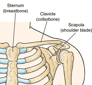
The clavicle, also known as the collarbone, is a long and slender bone that connects your sternum (breastbone) to your scapula (shoulder blade). It is one of the most visible and palpable bones in your body, and it plays a vital role in supporting and stabilizing your upper limb. Here, we will describe extensively the clavicle, focusing on two aspects: its articulation with the manubrium and scapula, and the muscles attached and their role in movement.
Surface Features of the Clavicle
The clavicle is an elongated, S-shaped bone that rests horizontally at the sternum across the upper part of the ribcage, and the acromial end of the scapula. It is an important part of the skeletal system since it plays an essential role in everyday functional movement, serving as the connection between the axial skeleton and the pectoral girdle. As a result, the clavicle is able to act as a brace for the shoulder, allowing weight to be transferred from the upper limbs to the axial skeleton.
The clavicle has three main functions:
- Attaches the upper limb to the trunk as part of the ‘shoulder girdle’.
- Protects the underlying neurovascular structures supplying the upper limb.
- Transmits force from the upper limb to the axial skeleton.

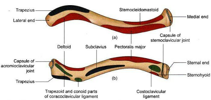
The clavicle can be divided into a sternal end, a shaft and an acromial end. The sternal end contains a large facet for articulation with the manubrium of the sternum at the sternoclavicular joint. The inferior surface of the sternal end is marked by a rough oval depression for the costoclavicular ligament, a ligament of the SC joint. The shaft of the clavicle acts as a point of origin and attachment for several muscles which include :deltoid, trapezius, subclavius, pectoralis major, sternocleidomastoid and sternohyoid. The acromial end houses a small facet for articulation with the acromion of the scapula at the acromioclavicular joint. It also serves as an attachment point for two ligaments: conoid tubercle – attachment point of the conoid ligament, the medial part of the coracoclavicular ligament. Trapezoid line – attachment point of the trapezoid ligament, the lateral part of the coracoclavicular ligament.
Articulation with the Manubrium and Scapula
The clavicle has two ends: the medial end and the lateral end. The medial end articulates with the manubrium, which is the upper part of the sternum. This joint is called the sternoclavicular joint, and it is the only bony connection between the axial skeleton (the skull, spine, and rib cage) and the appendicular skeleton (the limbs and girdles). The sternoclavicular joint is a synovial joint, meaning that it has a fluid-filled capsule that allows some movement. The movement of the clavicle at this joint is mainly elevation and depression (raising and lowering the shoulder), and protraction and retraction (moving the shoulder forward and backward).
The lateral end of the clavicle articulates with the acromion, which is a projection of the scapula. This joint is called the acromioclavicular joint, and it is also a synovial joint. The movement of the clavicle at this joint is mainly upward and downward rotation of the scapula, which affects the position of the glenoid cavity (the socket where the humerus, or upper arm bone, fits). The acromioclavicular joint also allows some gliding of the clavicle on the acromion.
The clavicle acts as a strut that keeps the scapula in a lateral position, away from the thorax. This allows the upper limb to have a greater range of motion and more freedom of movement. The clavicle also serves as a transmission of force from the upper limb to the axial skeleton, and vice versa. For example, when you push something with your arm, the force is transmitted from the humerus to the scapula, then to the clavicle, and then to the sternum. Conversely, when you shrug your shoulders, the force is transmitted from the sternum to the clavicle, then to the scapula, and then to the humerus.
Muscles Attached and Their Role in Movement
The clavicle provides attachment sites for several muscles that are involved in the movement and stabilization of the upper limb. These muscles are:
The trapezius, which is a large, triangular muscle that covers the back of the neck and the upper part of the back. The trapezius has three parts: the upper, middle, and lower fibers. The upper fibers attach to the lateral third of the clavicle, and they help to elevate and upwardly rotate the scapula.

The sternocleidomastoid, which is a thick, strap-like muscle that runs from the sternum and the medial third of the clavicle to the mastoid process of the skull. The sternocleidomastoid helps to flex and rotate the head and neck, as well as to elevate the sternum and the clavicle during inspiration (breathing in).
The pectoralis major, which is a large, fan-shaped muscle that covers the chest. The pectoralis major has two heads: the clavicular head and the sternal head. The clavicular head attaches to the medial half of the clavicle, and it helps to flex, adduct, and medially rotate the humerus. The sternal head attaches to the sternum and the costal cartilages of the ribs, and it helps to adduct and medially rotate the humerus.
The deltoid, which is a thick, triangular muscle that covers the shoulder. The deltoid has three parts: the anterior, middle, and posterior fibers. The anterior fibers attach to the lateral third of the clavicle, and they help to flex, abduct, and medially rotate the humerus. The middle fibers attach to the acromion, and they help to abduct the humerus. The posterior fibers attach to the spine of the scapula, and they help to extend, abduct, and laterally rotate the humerus.
The subclavius, which is a small, cylindrical muscle that lies under the clavicle. The subclavius attaches to the first rib and the inferior surface of the clavicle, and it helps to depress and stabilize the clavicle.
The clavicular head of the biceps brachii, which is one of the two heads of the biceps brachii, a large muscle that runs along the front of the upper arm. The clavicular head attaches to the apex of the coracoid process of the scapula and the medial half of the clavicle, and it helps to flex and supinate the forearm, as well as to flex the humerus.
As you can see, the clavicle is a very important bone for your upper limb, as it connects, supports, and moves it in various ways. The clavicle also gives shape and definition to your shoulder and neck, and it can be easily felt and seen under your skin.
Next on the list is the ossification of clavicle
The clavicle is a very interesting bone to learn about, as it has different ossification centers depending on its end. The lateral end of the clavicle is the first bone to start ossification at around 5th-6th weeks of gestation. It is also the last ossification center to fuse, around 22-25 years of age. The lateral end has intramembranous ossification, which means that the bone is formed by the deposition of bone matrix within the membrane that surrounds the cartilage.
The medial end of the clavicle, which articulates with the manubrium of the sternum, is the last bone to start ossification, around 15 years of age. It is also the first bone to fuse, as it forms part of the sternoclavicular joint, which is the only bony connection between the axial skeleton and the appendicular skeleton.
Fracture of the clavicle
The clavicle is particularly susceptible to fracture due to its relative size and role in transmitting forces from the upper limb to the axial skeleton. The most common mechanism of injury is a fall onto the shoulder or onto an outstretched hand³. With the clavicle arbitrarily divided into thirds: 15% of fractures occur in the lateral third, 80% occur in
the middle third, 5% occur in the medial third³. After a fracture, the lateral end of the clavicle is displaced inferiorly by the weight of the arm and displaced medially by the pectoralis major. The medial end is pulled superiorly by the sternocleidomastoid muscle.
Management of a clavicular fracture can be conservative (e.g. sling immobilisation) or operative (e.g. open reduction and internal fixation). The supraclavicular nerves lie in close proximity to
the clavicle and are occasionally sacrificed during a surgical repair – resulting in a numb patch over
the upper chest and shoulder.
Summary of this section !!
Here are some other important information to note about the clavicle bone:
- The clavicle is the only bone that connects the upper limb to the rest of the skeleton. It happens at the sternoclavicular joint, which is the only bony connection between the axial skeleton (the skull, spine, and rib cage) and the appendicular skeleton (the limbs and girdles).
- The clavicle is also the only long bone that lies horizontally in the human body. It acts as a strut that keeps the scapula (shoulder blade) in place so the upper limb can hang freely.
- The clavicle has different ossification centers depending on its end. The lateral end of the clavicle is the first bone to start ossification at around 5th-6th weeks of gestation. It is also the last ossification center to fuse, around 22-25 years of age. The lateral end has intramembranous ossification, which means that the bone is formed by the deposition of bone matrix within the membrane that surrounds the cartilage.
- The medial end of the clavicle, which articulates with the manubrium of the sternum, is the last bone to start ossification, around 15 years of age. It is also the first bone to fuse, as it forms part of the sternoclavicular joint, which is a synovial joint, meaning that it has a fluid-filled capsule that allows some movement.
References
(1) Clavicle | Skeleton of the upper limb | Upper Extremity. https://anatomy.app/article/skeleton-of-the-upper-limb/clavicle.
(2) Clavicle: Anatomy, Function, and Treatment - Verywell Health. https://www.verywellhealth.com/clavicle-anatomy-5089028.
(3) Clavicle | Definition, Anatomy, & Function | Britannica. https://www.britannica.com/science/clavicle.
(4) Clavicle: Anatomy, Function, and Treatment - Verywell Health. https://www.verywellhealth.com/clavicle-anatomy-5089028.
(5) Clavicle: Anatomy and clinical notes | Kenhub. https://www.kenhub.com/en/library/anatomy/the-clavicle.
(6) Clavicle | Radiology Reference Article | Radiopaedia.org. https://radiopaedia.org/articles/clavicle.
(7) Ossification and Clinical Features of the Human Clavicle. http://www.actforlibraries.org/ossification-and-clinical-features-of-the-human-clavicle/.
(8) Ossification centers of the pectoral girdle - Radiopaedia.org. https://radiopaedia.org/articles/ossification-centres-of-the-pectoral-girdle.
(9) Clavicle | Radiology Reference Article | Radiopaedia.org. https://radiopaedia.org/articles/clavicle.
(10) Ossification centers | Radiology Reference Article | Radiopaedia.org. https://radiopaedia.org/articles/ossification-centres?lang=us.
(11) The Clavicle - Functions - Landmarks - Fractures - TeachMeAnatomy. https://teachmeanatomy.info/upper-limb/bones/clavicle/.
(12) Clavicle | Complete Anatomy - Elsevier. https://www.elsevier.com/resources/anatomy/skeletal-system/appendicular-skeleton/clavicle/22586.
(13) Clinical Anatomy of Clavicle – Medchrome. https://medchrome.com/basic-science/anatomy/clavicle/.


I am a complete beginner who resides in Africa's Western Hemisphere. My name is James, but you may reach out to me through the Facebook page [James Kossy] (https://www.facebook.com/christ.messenger.904) Physics, chemistry, and biology are the three topics that I find most enjoyable. My current studies are taking place at the university level, with the intention of becoming a recognized professional in physiotherapy. I am fascinated by all things technological, and I take pleasure in contributing to the fascinating technological advancements that are taking place throughout the world today. In my spare time, I'd like to learn more about programming and help others with any technical problems they may be having. 💞 ***🌹❤️ Thank you so much to everyone who has supported me thus far. ****💞 At the moment, I don't have the right words to say how much I appreciate all of your help. You never cease to astonish me with your generosity. For me, this has turned into a haven of enjoyment. Thanks to colleagues like you, this has all been possible. You've been a great support for me. Everything you have done for me and my family has been greatly appreciated, and I will always be grateful to you. 💕.
Posted Using InLeo Alpha
Thanks for your contribution to the STEMsocial community. Feel free to join us on discord to get to know the rest of us!
Please consider delegating to the @stemsocial account (85% of the curation rewards are returned).
You may also include @stemsocial as a beneficiary of the rewards of this post to get a stronger support.