Introduction
We follow my dear readers with the wonderful analysis of all those rays invisible to our eyes, and which structure a great part of our essential electromagnetic spectrum, where we start with our referential fraction, that is, the white or visible light and such spectral fraction constitutes a minimum expression of the mentioned electromagnetic spectrum. However, through the knowledge obtained from the white light, man has managed to structure the other spectral fractions even though they are imperceptible to our optical systems.
Therefore, we can say that until now we have analyzed not only the fraction of our white or visible light, but also other spectral fractions such as ultraviolet rays and also infrared rays, of these rays we knew some of their essential applications both in the scientific-technological world and with it in different activities related to the development of our lives, in all senses.
In this opportunity we will analyze in a general way to the recognized X-rays, and the same as the ultraviolet and infrared are invisible to our natural optical systems (eyes), nevertheless, this type of electromagnetic radiation has great utility between the human beings above all in the area of the medicine and consequently in the care of our health in spite of the fact that the same ones belong to the family of ionizing radiations, this aspect we will continue extending it later.
It is important to emphasize that since the beginning of the structuring of the electromagnetic spectrum we have relied on the principles offered by geometric optics, among which we find its valuable conceptualization of light rays whose propagation is rectilinear, in addition to other aspects or phenomena studied by geometric optics such as reflection, refraction and diffraction, all this also supported by principles provided by physical optics and their wavelengths.
The important thing of the study of these spectral fractions is that it has allowed us to use these rays in different applications, but also we have been able to learn how to protect ourselves from them, very important aspects since in spite of not being ionizing like the ultraviolet (UV) and infrared (IR) rays a long exposure to them can generate irreversible damages in our skin.
We must point out that the main natural source of emission of electromagnetic radiation is our central star, that is, the Sun, where we can find the powerful gamma rays of short wavelength but high frequency to infrared radiation with longer wavelength but lower frequency, the above described allows us to express that in that interval we can get to X rays (R-X), because its spectral fraction is between gamma and ultraviolet rays.
Therefore, the X rays are constituting our electromagnetic spectrum and whose wavelength is less than that belonging to the white or visible light, this leads us to highlight the use of any machine, device or tool created by man to capture and see the action of X rays, in this way is that we have been able to see images of the interior of our body thanks to the action of these rays on certain material photosensitive to them and the same we know as radiographs.
In addition to the positive applications of X-rays, it will always be important to point out the risks when we are exposed to these X-rays in an uncontrolled way, because their main property or characteristics is that they belong to the family of rays or ionizing radiation and thus have the ability to change the electrical charges belonging to the atoms that make up the molecules that structure our body.
With the help of our referential fraction and its capacity of penetrability, we will be enjoying a wonderful home practice by means of the visible luminous rays of a lantern and placing it in contact with our palm and evaluating its power of penetrability in the same one, this with the purpose of comparing this action with the effect generated by the x-rays when entering in contact with our body or any other matter.
X-Ray
In the same way as we did for ultraviolet (UV) and infrared (IR) rays, on this occasion we will use the elemental principle provided by physical optics, i.e., that the vital phenomenon of light is a type of electromagnetic radiation, and that is why we have called it radiant energy, without forgetting the fundamental principle of geometric optics, i.e., its conceptualization of a light ray whose propagation has a straight path.
It is important to make a small historical review and to express that the X rays (R-X) were discovered at the end of century XIX, very specifically in the year of 1895 by the recognized German physicist Wilhelm Conrad Roentgen, and was who I place the name to them of X rays (for being the X used generally to an unknown one) by not knowing initially its origins, nevertheless, for 1912 is managed to specify the nature of this radiation, through the phenomenon of the diffraction in crystals.
In an artificial way these rays are managed to originate when we make that a certain beam of accelerated electrons (voltage of thousands of volts) collides with a receiving material (tungsten), when being generated this shock with the atoms of the receiving matter, these electrons are going to be slowed down causing this way that the same ones dissipate part of their energy, and with this loss of energy, a fragment (almost the totality) was transformed in heat, and consequently the rest in electromagnetic radiation, in this case X rays, as we can visualize in the following figure 1.
Figure 1. X-ray generation
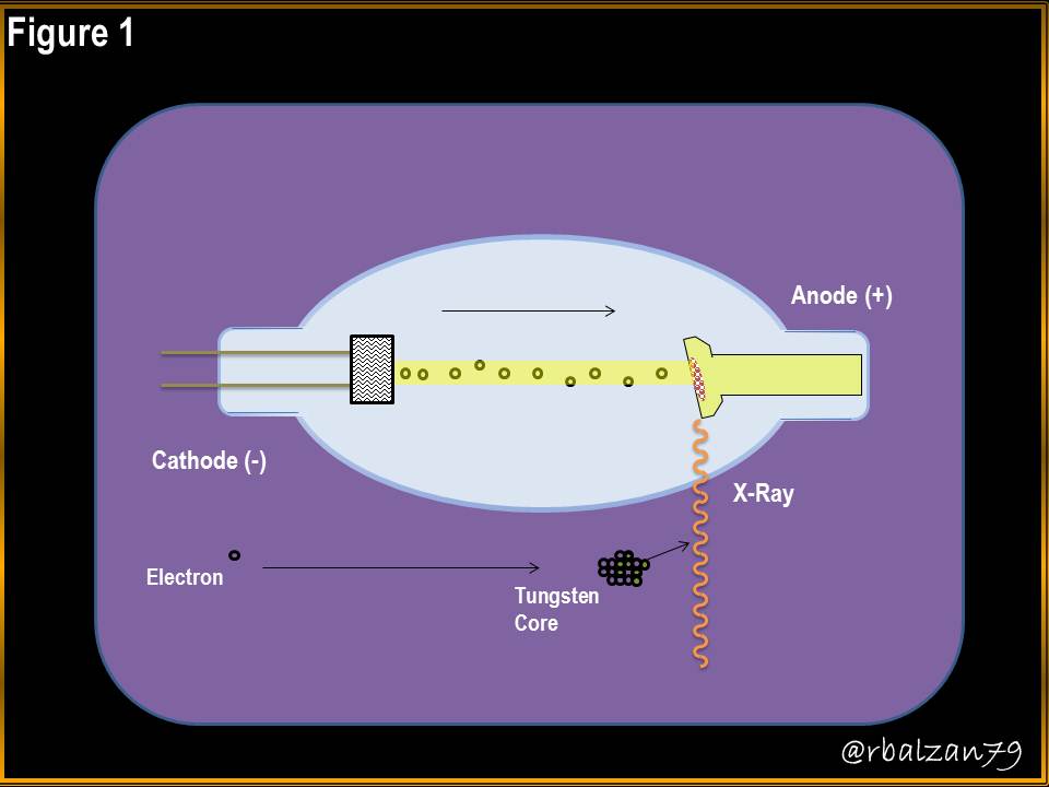
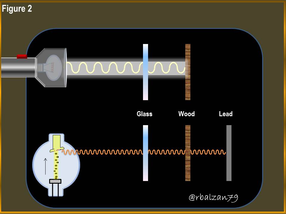
This type of electromagnetic radiation differs from our referential spectral fraction (white or visible light) in its wavelength and frequency, because X-rays (R-X) have shorter wavelengths and thus higher frequency capacity, and thus their range of action as already expressed, is located between ultraviolet and gamma rays, this short wavelength allows the x-rays to have greater energy capacity than white or visible light, this outstanding aspect allows it to go through our body, as well as other materials that white light cannot achieve, as we can visualize in the following figure 2.
Figure 2. X-ray, and its outstanding penetrability characteristic
Therefore, the X rays when possessing this high energy amount makes them belong to the family of ionizing electromagnetic radiations, and due to this when these rays interact with some matter they will lose certain amount of energy with which they can get to separate electrons that conform the atoms of the molecules of this matter, and this way the ions are originated, then, in more practical terms we can express that the ionization process consists of starting an electron to a determined electrically neutral atom.
According to the above, we must be very careful when our body is exposed to this type of electromagnetic radiation because of the ionizing characteristic described above, that is, the penetrating power of X-rays.
It is important to be able to observe the power of penetrability of white light in our body through the implementation of a flashlight, for this we will make a small but significant practical experience and verify if with the white light impacting on any part of our body is possible to visualize the interior, so it would be interesting to ask:
Can light rays have any penetrability in any part of our body, and if so, can they be compared with those of X-rays?
Next we will present the steps of our practical experience realized with materials of easy access in our houses, this will be able to visualize it in the following images:
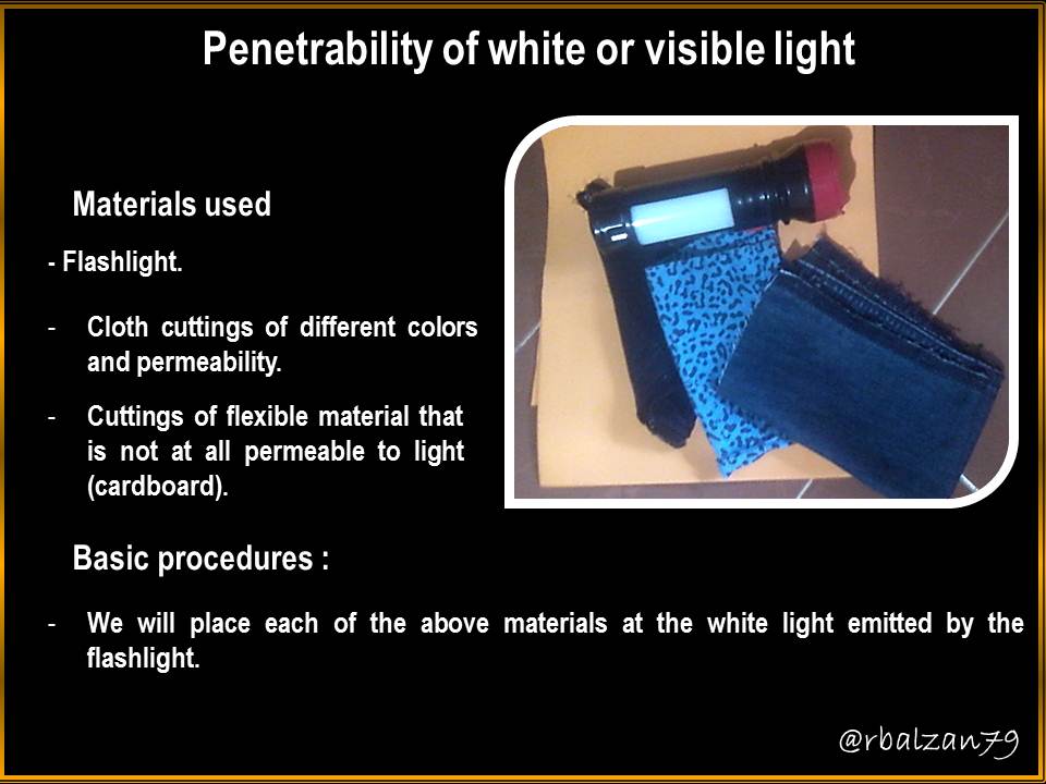
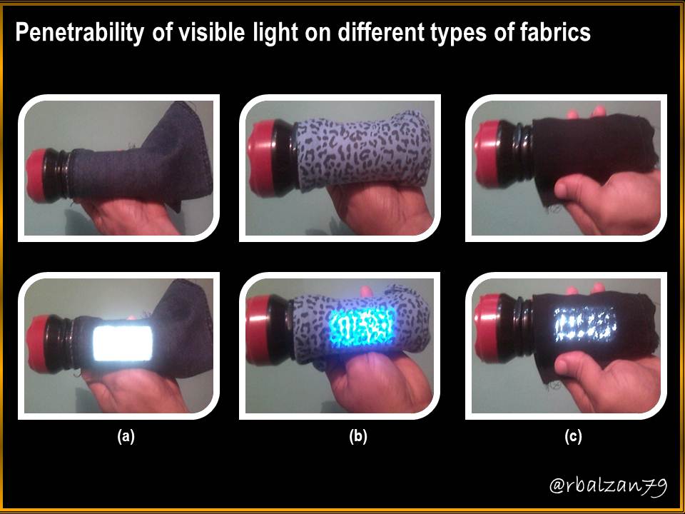
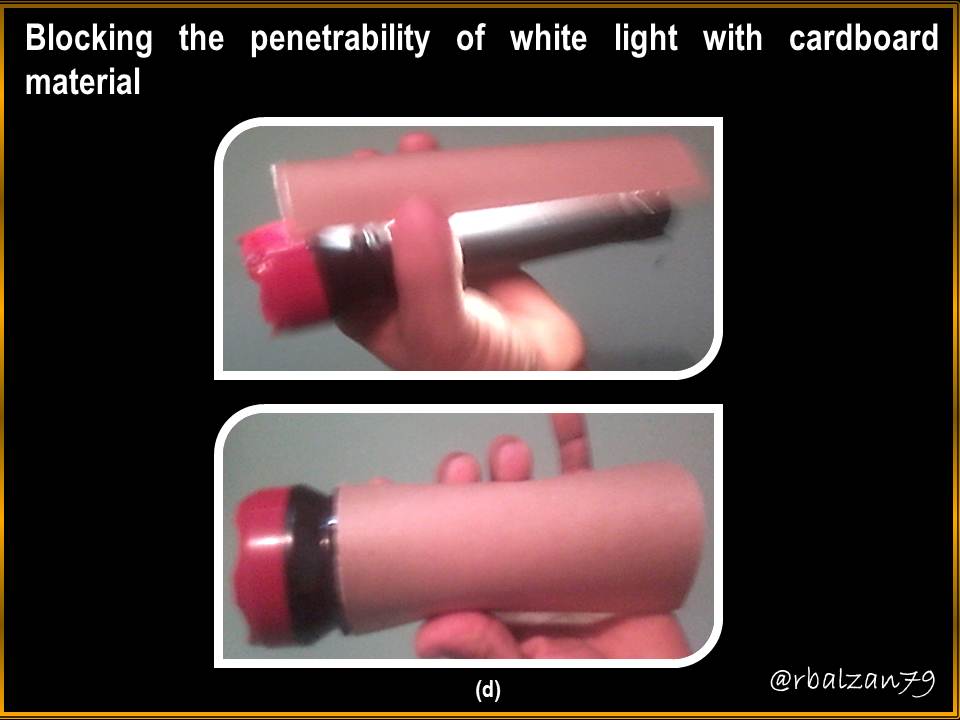
Once our home experience was made, in the images they could notice that the light rays visible to our eyes managed to penetrate in different types of fabrics as it is observed in the figures (a), (b) and (c), when we apply this light rays in another type of material as the cardboard the penetrability is null as it is observed, that is to say, that it is annulled, as we can observe in the figure (d).
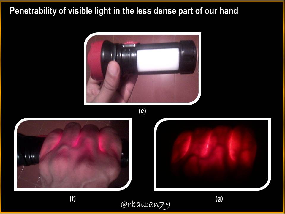
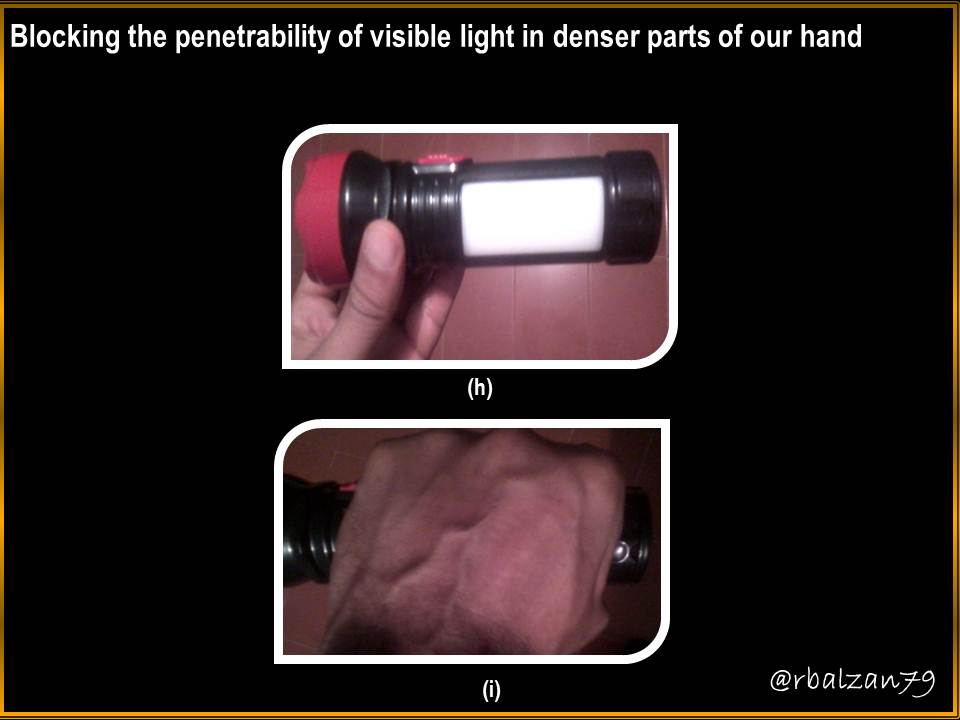
Then we take the light rays to a part of our palm (g) and observe that we can visualize part of the interior of it, however, this characteristic is reduced or annulled when we move the light to a denser part of the hand, and therefore, the effect of penetrability is lost (i).
To culminate with the analysis of the referred practice, we can give answer to the raised question, and although the white light could penetrate in some part of our hand, this capacity is lost or is annulled when we transfer the rays of light towards a more dense part of our hand, and with it we demonstrate that the visible light does not possess the same capacity of penetrability of the x-rays, since the x-rays possess major energetic capacity and consequently major capacity of penetrability up to coming to be ionizing, as they observed in the figure 2.
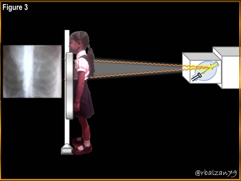
There are many applications of X-rays, but we must emphasize that one of the most important is in the area of medicine, that is, medical X-rays, through radiographs as we can see in the following figure 3.
Figure 3. X-ray
X-ray spectrum
This type of radiation is electromagnetic and invisible to our natural optical systems (eyes), in the same way as ultraviolet (UV) and infrared (IR) rays, this means that these rays have certain wavelengths with rectilinear propagation as established by physical-geometric optics, the spectral fraction of these X-rays can be seen in the following figure 4.
Figure 4. X-ray spectrum
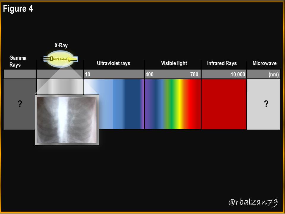
In the previous figure 4, they could observe the spectrum of the X rays, the color of this radiation can vary at the moment of the impact with a certain matter of our universe, we use the color white-gray by the importance of the developed to black and white of the photosensitive plates (radiographies) to these X rays, nevertheless, through the observation with a telescope of X rays in the astronomical world, other colors can be visualized.
Conclusion
With the present article, we continue with the conformation of our essential electromagnetic spectrum, where, we have already analyzed our referential spectral fraction, that is to say, the white or visible light, from there we pass with the interesting analysis of the invisible radiations to our glance, and this way we have found with the infrared rays (IR), ultraviolet (UV) and in this opportunity with the recognized X rays.
This type of radiation has short wavelengths which represents a higher frequency than the previous analyzed radiations, this gives it a great energetic capacity becoming ionizing when impacting with certain matter due to its great power of penetrability, this aspect of penetrability allowed us to carry out a small practical experience, where, we used an artificial source of white light to observe the power of penetrability of the white light in some part of our palm.
In spite of the fact that the white light was able to penetrate the interior of one part of our palm, it was not able to penetrate through another part of our palm with a greater density of material, and in addition, when using other materials such as cardboard, it was not able to pass through it, or at least at first sight, we were not able to observe this capacity of penetration.
We end up concluding that in spite of this specific penetrability in our palm, it is not possible to compare this capacity of penetrability with the one offered by the X-rays, due to the fact that the same one possesses a greater frequency and with it a greater energetic quantity than that of the white or visible light, in figure 2 they could observe the relation of penetrability of these two electromagnetic radiations, where, the X-rays manage to overcome the visible light.
Until another delivery my appreciated readers of Hive.blog, specially to the members of the great community of #Stemsocial, which receives the support of another wonderful community like #curie, reason why I recommend widely to be part of this exemplary project, since they allow us to emphasize the wonderful task of the academy and the enormous work of all the field of science.
Note: Some images were elaborated by means of Power Point, and the animated gif was elaborated with the application of PhotoScape, the photostatic images were captured by the optical instrument (camera) of the cell phone ZTE BLU Life Play 2.
Bibliographic References
[1]Charles H. Lehmann. Geometría analítica
[3]X-Rays