Contraction in the simplest sense is shortening of a muscle fibre. When muscles receive stimulation from the nervous system, the required fibres simultaneously shorten to produce a contraction of the overall muscle. It seems simple enough, right? However, the mechanisms by which muscles are able to shorten is remarkably complicated.
How do muscle fibres shorten?
To understand how muscles are able to shorten, we must look at their ultrastructure, which is shown in figure 1 below.
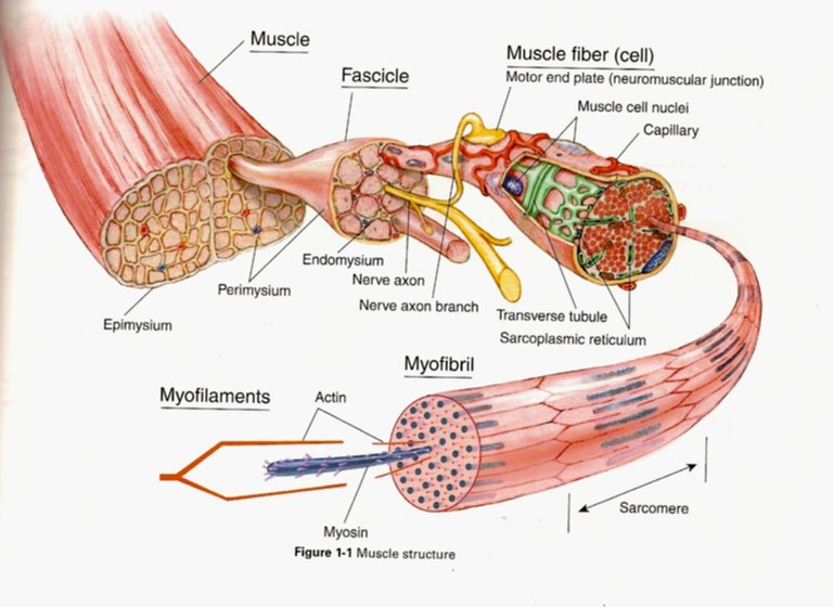
figure 1. Ultrastructure of skeletal muscle
The contractile unit of muscle is called a sarcomere. It is the shortening of each individual sarcomere which results in the shortening, and thus contraction of muscle at a macroscopic level.
figure 2. Sarcomere ultrastructure
During a contraction the size of a sarcomere will decrease, bringing the Z lines (shown in figure 1) closer together. This is achieved through the interaction between actin and myosin.
Myosin is known as the thick filament within muscle and actin the thin filament respectively. During a muscle contraction, myosin heads bind to the thin actin filament. This causes a conformational change in the myosin heads, resulting in the myosin heads flicking inwards towards the M-line (figure 2). This causes the sarcomere to shorten. For clarity, this is shown in the animation below.
figure 3. Sarcomere shortening
ATP (which is used as an energy currency within cells) is then used to detach the myosin heads from their actin binding sites. Myosin can bind again to the binding sites on the thin filament for a continual contraction. It is interesting that energy is required to detach the myosin heads from actin after a contraction, rather than to initiate a contraction itself. This specific role of ATP is the reason muscles become rigid and tight after death in a process called rigor mortis. Following death, stores of ATP in the body are used up, resulting in myosin heads remaining bound to actin binding sites within muscles, giving a rigid state characteristic of rigor mortis.
Obviously, all the muscles in your body do not all contract simultaneously. This means there must be a mechanism that regulates when myosin heads are able to bind to actin filaments.
The solution to this problem is achieved by using calcium (Ca2+).
It makes sense that the rested state of muscles is to be relaxed. Therefore, under normal circumstances myosin heads are not able to bind to actin and no contraction will occur. This is accomplished because the binding sites are blocked by the protein tropomyosin. The tropomyosin itself is attached to another regulatory protein called troponin.
Upon the initiation of a muscle contraction Ca2+ is released and binds to troponin. This causes a conformational change which results in tropomyosin moving away from actin to reveal the myosin head binding sites, thus allowing a muscle contraction to occur. When a muscle is required to stop contracting the Ca2+ is simply removed from troponin to allow tropomyosin to block the binding sites once again.
How is Ca2+ released?
The Ca2+ released to bind to troponin to cause a muscle to contract is stored in a specialised organelle within skeletal muscle known as the sarcoplasmic reticulum. When a nervous impulse arrives at a muscle cell giving the information to contract, this electrical signal is sent to the sarcoplasmic reticulum down a process within muscle cells called the transverse (T) tubule. The arrival of this impulse causes Ca2+ to be released from the sarcoplasmic reticulum and to bind to troponin.
Stay strong, we have reached the end! Finally we know how muscles initiate, sustain and terminate a contraction. It seems such a complicated process, for simple actions that we are able to achieve even without thought.
Here, we have only discussed skeletal muscles. The physiology that allows the contraction of cardiac muscle is similar and also requires actin and myosin. However, smooth muscle contraction is more complicated and many of its mechanisms are yet undiscovered.
References:
Rigor mortis
Mechanism of muscle contraction
Troponin and tropomyosin
@originalworks
The @OriginalWorks bot has determined this post by @ovij to be original material and upvoted it!
To call @OriginalWorks, simply reply to any post with @originalworks or !originalworks in your message!
To nominate this post for the daily RESTEEM contest, upvote this comment!
For more information, Click Here!
This post recieved an upvote from minnowpond. If you would like to recieve upvotes from minnowpond on all your posts, simply FOLLOW @minnowpond
Incredible detail! Great job...
Thanks man, really appreciate it!
Hey @ovij I have been meaning to tell you this for a while. I really enjoy your blog! :)
Thank you very much, yours too. Especially your latest post on mental abnormality and creativity.
Good luck dear👍👍👍
Congratulations! This post has been upvoted from the communal account, @minnowsupport, by justtryme90 from the Minnow Support Project. It's a witness project run by aggroed, ausbitbank, teamsteem, theprophet0, someguy123, neoxian, followbtcnews/crimsonclad, and netuoso. The goal is to help Steemit grow by supporting Minnows and creating a social network. Please find us in the Peace, Abundance, and Liberty Network (PALnet) Discord Channel. It's a completely public and open space to all members of the Steemit community who voluntarily choose to be there.
This post has received a 0.78 % upvote from @drotto thanks to: @banjo.
upvoted my brother
I love me some anatomy, especially when it is about muscles!!!