Introduction
For the purpose of a better understanding, consider the brachial plexus a circuit of wires connecting the upper limb to the power source (the Central Nervous System). It is anatomically considered as a network of nerve fibers that innervate the skin and muscles of the upper limb and is made up of the anterior rami(divisions) of the last four cervical nerves, C5, C6, C7, C8 and the first thoracic nerve, T1. It transfers actions as authored by the Central Nervous System(CNS) to be carried out by the upper limb and conveys information from the upper limb to be interpreted by the CNS( Brain and spinal cord).
In the body, four major nerve plexuses can be found in different locations and having different functions. With their names depicting the areas in which they have a main function, they include:
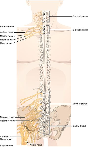 The Major Nerve Plexuses in the Body (License: CC-BY 4.0, Author: OpenStax]:
Wikipedia Commons
The Major Nerve Plexuses in the Body (License: CC-BY 4.0, Author: OpenStax]:
Wikipedia Commons - Cervical plexus
- Brachial plexus
- Lumbar plexus
- Sacral plexus
To the vast majority, the arm, hand and upper limb mean one and the same thing, but anatomically, these are different terms entirely. I believe a knowledge of this will help give a better understanding on what is about to be discussed.
- The arm is the region between the shoulder joint and the elbow joint.
- The forearm is the region between the elbow joint and the wrist while,
- The hand is the region from the wrist down to the phalanges(fingers)
 The Brachial Plexus Showing its Component Segment and Branches (License: Public Domain]:
Wikipedia Commons
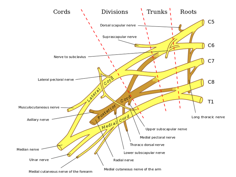
The Brachial Plexus Showing its Component Segment and Branches (License: Public Domain]:
Wikipedia Commons As a matter of fact, the brachial plexus is made up of:
- Roots
- Trunks
- Divisions
- Cords
Roots
At every vertebral level, spinal nerves leave the intervertebral foramina and divide into two; the anterior and posterior rami. The anterior rami of C5, C6, C7, C8 and T1 are what form the roots of the brachial plexus. They unite to form the trunks while the posterior rami go on to supply the skin of the trunk.
Trunks
At the base of the neck, the roots join to form three distinct trunks.
- C5 and C6 join to form the superior trunk
- C7 continues as the middle trunk
- C8 and T1 form the inferior trunk.
Each trunk then divides into anterior and posterior parts and unite to form cords. I truly hope that the nature of division is not getting confusing as it is important if one must understand how the nerves that innervate a muscle or part of skin is formed. This helps to know the part of the brachial plexus that is damaged when a particular muscle or set of muscles starts to malfunction.
Cords
Once the trunks divide, they unite to form the cords in this order:
- The anterior divisions of the superior and middle trunks form the lateral cord
- The posterior division of all the trunks unite to form the posterior cord
- The anterior division of the inferior trunk continues as the medial cord.
These cords are named in respect to their relation to the axillary artery. They give rise to five major terminal branches in the axilla and the proximal part of the arm.
Terminal branches
The five major branches include(from lateral to medial):
- Musculocutaneous nerve
- Axillary nerve
- Radial nerve
- Median nerve
- Ulnar nerve
Musculocutaneous nerve
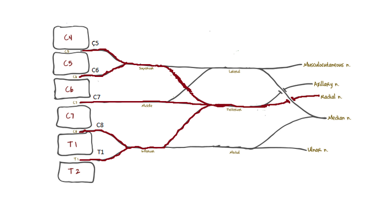
) Image Showing Composition and Formation of the Musculocutaneous Nerve (License: CC-BY-SA 3.0, Author: Chris Tablot]:
Wikipedia Commons
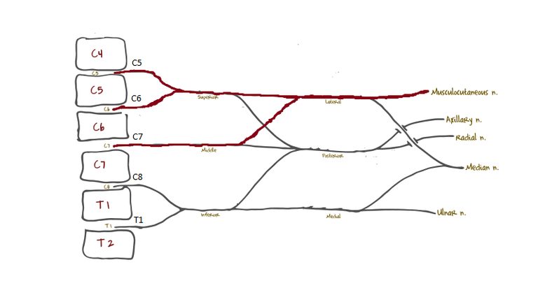
Image Showing Composition and Formation of the Musculocutaneous Nerve (License: CC-BY-SA 3.0, Author: Chris Tablot]:
Wikipedia Commons This nerve emerges as the terminal branch of the lateral cord of the brachial plexus in the axilla(arm pit), enters the arm, pierces a muscle(the coracobrachialis muscle) and continues down between two other muscles(the brachialis and biceps muscles). It terminates by going superficial to become the lateral cutaneous nerve of the forearm .
It has root values of C5, C6 and C7.
- motor function:
It supplies all three muscles of anterior part of the arm(biceps brachii, coracobrachialis, brachialis). These muscles flex the arm at the shoulder and elbow joint and the biceps supinates the forearm at the elbow joint. These muscles collectively give the upper limb the ability to lift a tea cup from the table for a drink.
- sensory function:
It innervates the skin over the lateral part of the forearm(it carries sensory information to the brain from this part).
) Image Showing Composition and Formation of the Axillary Nerve (License: CC-BY-SA 3.0, Author: Chris Tablot]:
Wikipedia Commons
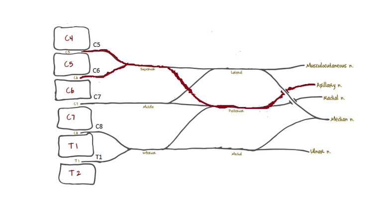
Image Showing Composition and Formation of the Axillary Nerve (License: CC-BY-SA 3.0, Author: Chris Tablot]:
Wikipedia Commons Axillary nerve
This nerve emerges as the terminal branch of the posterior cord of the brachial plexus, exits the axilla(arm pit) posteriorly through the quadrangular space and winds around the surgical neck of the humerus deep to the deltoid(the shoulder muscle).
It has root values of C5 and C6
Motor function :
It supplies the deltoid and teres minor muscles. The deltoid initiates movement during abduction(lifting sideways) of the arm and the teres minor aids in stability of the shoulder joint. The teres minor is a member of a group of four muscles that stabilize the elbow joint by attaching around the humeral head; the rotator cuff muscles.Sensory function :
It supplies the skin overlying the regimental badge area. The area just inferior to the shoulder muscle(deltoid)
Radial nerve
) Image Showing Composition and Formation of the Radial Nerve (License: CC-BY-SA 3.0, Author: Chris Tablot]:
Wikipedia Commons
Image Showing Composition and Formation of the Radial Nerve (License: CC-BY-SA 3.0, Author: Chris Tablot]:
Wikipedia Commons It has root values of C5-T1.
Motor function:
It innervates the muscles of the posterior compartment of the arm and forearm. These muscles help in stretching forth the upper limb, as in receiving somethingSensory function:
It innervates the skin of the lateral aspect of the arm below the deltoid, the skin of the posterior aspect of the arm, a strip of skin along the posterior aspect of the forearm and also innervates the posterior part of the lateral three and a half digits and parts of the palms related to them
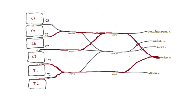) Image Showing Composition and Formation of the Median Nerve (License: CC-BY-SA 3.0, Author: Chris Tablot]:
Wikipedia Commons
Image Showing Composition and Formation of the Median Nerve (License: CC-BY-SA 3.0, Author: Chris Tablot]:
Wikipedia Commons Median nerve
The median nerve is a combination of the terminal branches of the lateral and medial cords of the brachial plexus. on entering the arm, it runs laterally till it gets to the middle of the arm where it crosses over to the medial aspect and descends into the cubital fossa. It enters the forearm from here and travels between the extensor digitorum profundus and extensor digitorum superficialis. It then enters the hand by passing through the carpal tunnel(a passage for structures passing from the forearm to the hand and from the hand to the forearm).
It has a root value of C6-T1
Motor function:
It supplies all muscles of the anterior compartment of the forearm except the ulnar head of the flexor carpi ulnaris and the medial part of the flexor digitorum profundus. These muscles cause pronation of the forearm and flexion of the wrist and digits of the hand. It also innervates the thenar muscles and the two lateral lumbricals. The muscles innervated by this nerve aids in curving the hand as in making a ‘snake head’ sign.sensory function:
It innervates the lateral part of the hand and the anterior surface of the lateral three and a half digits.
Ulnar nerve
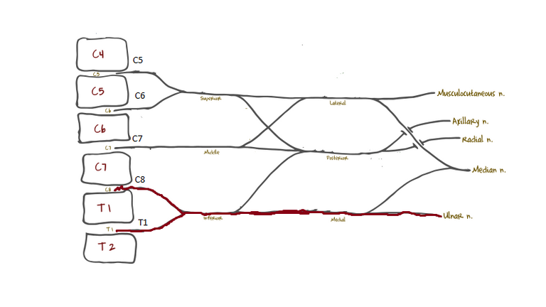) Image Showing Composition and Formation of the Ulnar Nerve (License: CC-BY-SA 3.0, Author: Chris Tablot]:
Wikipedia Commons
Image Showing Composition and Formation of the Ulnar Nerve (License: CC-BY-SA 3.0, Author: Chris Tablot]:
Wikipedia Commons Motor function:
Supplies the ulnar head of the flexor carpi ulnaris, the medial part of the flexor digitorum profundus and the hand muscles except the thenar muscle and the two lateral lumbricals. The muscles innervated by this nerve nerve help to grip a paper placed between two finger(adduction and abduction of the phalanges).Sensory Function:
Innervates the anterior and posterior surface of the medial one and a half fingers and the area of palm associated with them
Other branches given off at different points of the brachial plexus include:
At the root level
- dorsal scapular nerve
- long thoracic nerve
At the trunk level
- suprascapular nerve
- nerve to subclavius
From lateral cord
- lateral pectoral nerve
From medial cord
- medial pectoral nerve
- medial cutaneous nerve of arm
- medial cutaneous nerve of forearm
From posterior cord
- superior subscapular nerve
- thoracodorsal nerve
- inferior subscapular nerve
Clinical significance
- Cervical rib
this is an abnormally present rib which is present above the normal first rib, and arises from the seventh cervical vertebrae. this becomes a problem to the brachial plexus when it compresses its lower trunk.
- Prefixed and postfixed plexus
the normal root values of the brachial plexus as stated before is from C5-T1. In a situation where the first cervical contribution is from C4 and the last is from C8(C4-C8), it is known as a prefixed brachial plexus and when the first contribution is from C6 and the last from T2(C6-T2), it is called a post fixed brachial plexus.
- Erb’s palsy
This is otherwise called upper brachial plexus injury. it occurs due to a stretch or a tear of the upper nerve roots(C5 or C6) of the brachial plexus due to increase in the angle between the neck and shoulder. this causes a malfunctioning of the muscles supplied by the nerves with the contribution from these nerve roots(musculocutaneous nerve, axillary nerve, median nerve, radial nerve).
- Klumpke’s palsy
This is otherwise called lower brachial plexus injury. it occurs as a result of an excessive increase in the armpit angle(angle between the arm and the trunk). this causes a malfunctioning of the muscles supplied by the nerves with a contribution from T1(ulnar and median nerves).
Summary
From this post, one could tell how perfectly useless and immovable the upper limb would be if there wasn’t a brachial plexus. The brachial plexus is what brings life and function to the upper limb. It helps in the coordinated activities carried out by the limb and is also the main informant to the brain and spinal cord on matters concerning the upper limb as it provides a passageway for sensory information to pass through to them. Thus, an understanding on how these nerves control each of the muscles would be of great help in tracing accurately and easily what could be the cause of any malfunction.
Furthermore, any injury to the brachial plexus leads to the inability of the affected limb to carry out its functions as its supposed to, thus pointing more to the fact that the brachial plexus is one of the most important components of the upperlimb.
References
Moore, Keith L., Dalley, A. F., & Agur, A. M. R. (2010). Clinically Oriented Anatomy (6th ed.). Philadelphia: Lippincott Williams & Wilkins.
Drake, Richard; Vogl, A. Wayne; Mitchell, Adam W. M. (2009). Gray's Anatomy for Students E-Book. London: Churchill Livingstone
Sinnatamby, Chummy S., and R. J. Last. 1999. Last's anatomy: regional and applied. Edinburgh: Churchill Livingstone.
Chaurasia's. Human Anatomy: Regional and Applied Dissection and Clinical. 4th ed. New Delhi: CBS publisher and Distributors
Image sources
All images are from wikicommons licensed under creative commons and eligible for commercial use.
Thanks for reading