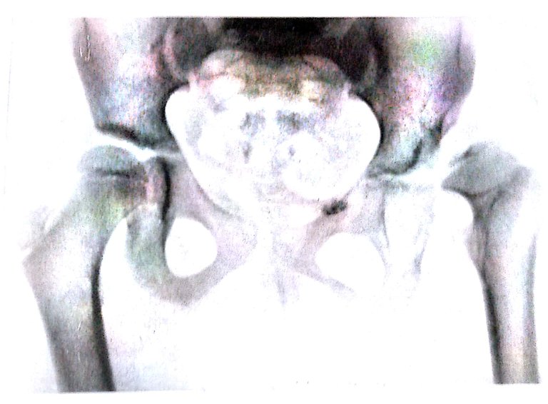
 Radiograph of the hip-joints of a girl age 6, showing stunted development of the head and neek of the left femar following unsuccessful attempts to reduce a congenital dislocation.
 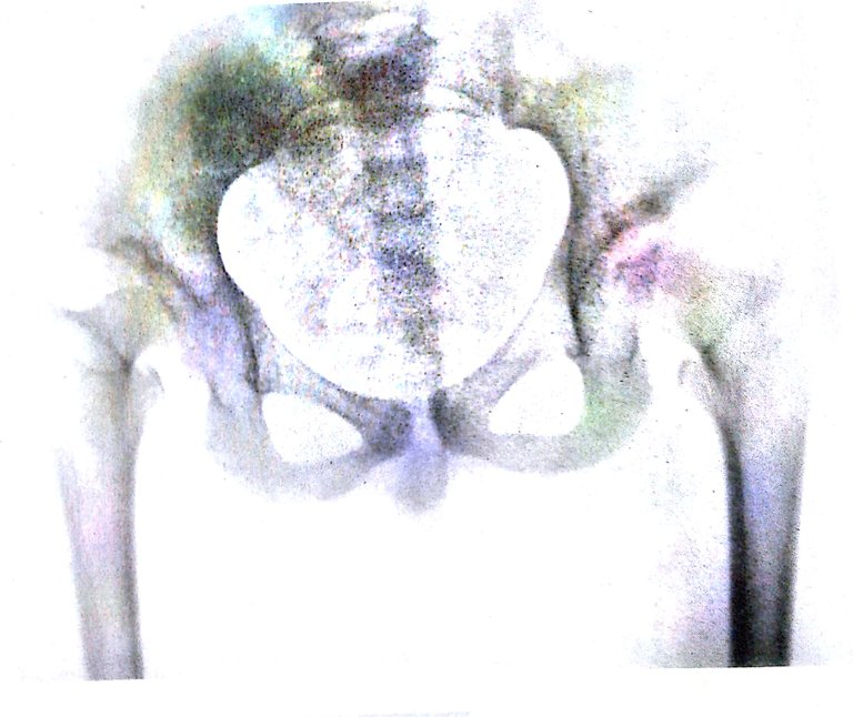 Radiograph of the hip-joints of a girl aged 13 years,showing bilateral stunted development of the femoral heads and neeks, though the bilateral dislocations had been completely reduced.
 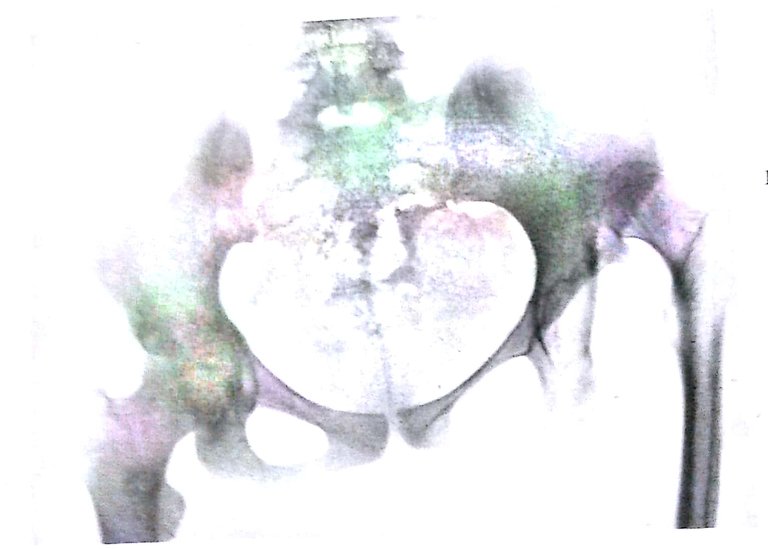 Radiograph of a girl aged 15, showing a congenital dislocation of the left hip-joint, Note the femoral head, neck and shipt are not so well developed as the right, and that afalsc aeetabulum has been developed in the left ilium.
Radiographs also show that when an epiphysis has been displaced by trauma, portions of the metaphysis are also displaced with it, i.e., it is not a clean separation through the metaphysis as we see in slipped epiphsis. In some cases of scurvy epiphyses are displaced as a result of separation through the decalcified zone in the diaphyses. The growth cartilage in these cases remains intact.
nice post
Helpful post
awesome
VERY NICE POST :)