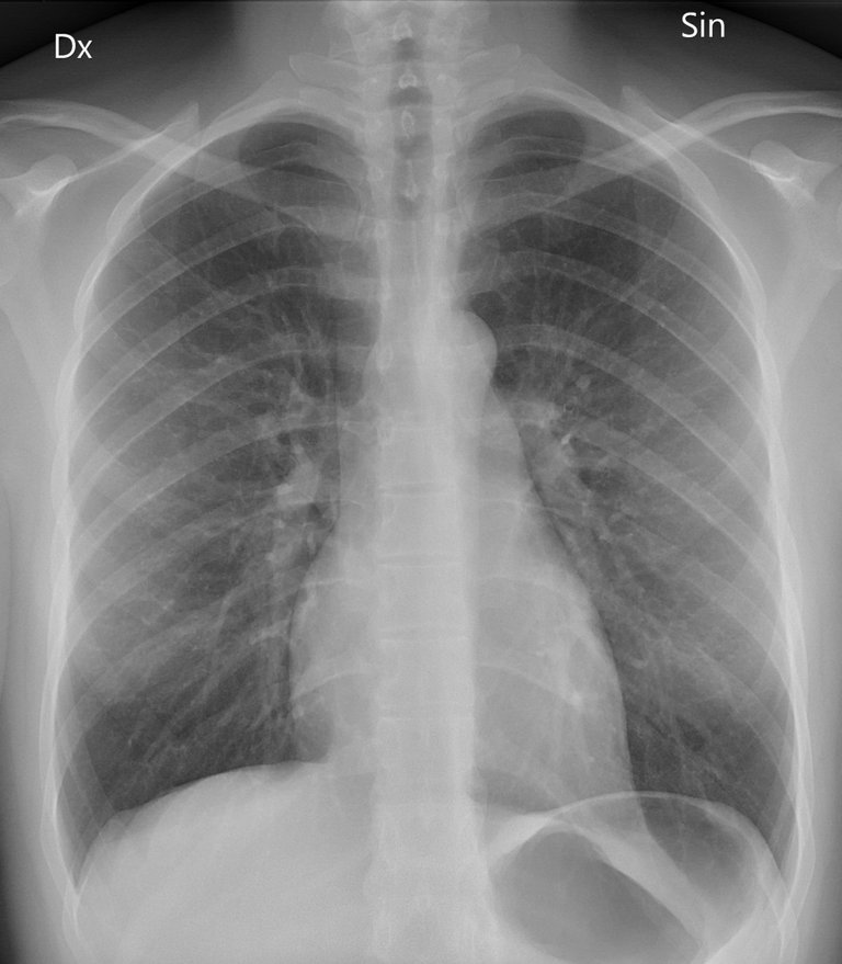
hey guys, welcome to Part-II of the article. In part-I, we discussed the basics of reading a normal chest X-ray. Now, in Part-II, we will discuss the abnormal findings in the chest X-ray. I explained the "ABCDE" system in my previous article. For those who are not familiar with this system, let me give you a hint. "ABCDE" system means what we should look for in an X-ray in a systematic way. it stands for Airways, Bones, Cardiac, Diaphragm, and Effusions. Keep in mind that, this system is only for a Chest X-ray. In this article, we will particularly look for the abnormal findings in the airways and bones.
The aim of this article, to give you a basic idea of what pathologies you can find in the airways and bones in an X-ray and how you can assess them correctly. so, let start the topic with the Airways.

Image: Chest Xray (Credits, Wikimedia, Reuse license)
Abnormalities in the Airways.
when we talk about the airways, what are the parts that we should look for in a chest X-ray? well, I covered this question briefly in my previous article. Those structures that you must look for are mainly, Trachea, left bronchus, and right bronchus. These structures are clearly visible and can be easily interpreted. Different abnormalities that you can look for are:
- Deviation of the Airway
- Foreign bodies in the Airway
- Blockage
- Narrowing of the Airways.
Deviation of the Airways:
We use "Trachea" as a reference to look for any type of deviation. This deviation might be the result of unequal intrathoracic pressure between the right and the left lung. There are various reasons that can lead to a change in intrathoracic pressure. some of them are Pneumothorax(air in the pleural space), atelectasis, effusions, or cancers.


Here in the picture given above, you can compare the features between normal and abnormal X-rays. Since the intrapleural pressure is always lower than the atmospheric pressure, once the air starts accumulating, it keeps going on. This might be because of accident or trauma that let the air in. Now, when the air starts accumulating, the intrapleural space starts to enlarge. After some time, it will enlarge enough to push the lung, mediastinum, and airways to the contralateral side. This results in the tracheal deviation to the contralateral side. That means it is pushed away from its original location. This phenomenon can be clearly seen in the above-mentioned image. See, how the lower left lung is collapsed due to pneumothorax which in turn is pushing the cardiac parts and the trachea to the contralateral side(right side in this case).
The pneumothorax pushes the structures away, but there are some conditions that pull the structures to the same side. Let's see some of the examples.
| Pushing | Pulling |
|---|---|
| Pneumothorax | Atelectasis |
| Cancer | Pulmonary Fibrosis |
| Effusion | Pleural Fibrosis. |
Foreign Bodies in the Airway.

This condition occurs when a foreign body enters the airways and causes choking. It can enter through the mouth, to the esophagus and trachea, blocking the airways. Most of these cases are recorded in small children between the age of 3 months to 6 years. They have a tendency to put small objects in the mouth. Thus, they enter the airways causing aspiration. In adults, it's very rare to do it intentionally, but still many cases of choking are seen.
In the given X-ray of a child, you can clearly see the foreign body lodged in the airway. Note that, this object is radio-opaque so we can visualize it clearly, but most of the foreign bodies are radiolucent, which makes it harder to visualize it properly. Therefore, an experienced radiologist is always preferred to interpret it. Now of course this condition can lead to some serious complications like atelectasis, fibrosis, bronchospasm, etc.
Narrowing of Airways

Image: Narrowing of the trachea(Credits, Wikimedia, reuse license)
Narrowing of airways is very easy to diagnose clinically and rarely requires an X-ray. But there are some critical cases where it is required. The narrowing can be seen during the inflammation of the trachea leading to subglottic stenosis. This inflammation of the airway is also known as acute laryngotracheobronchitis or Croup. This is just an example that causes the narrowing of the airway. Keep in mind that there can be many other reasons leading to this condition.
In the given Xray, we can see the narrowing of the trachea. Now, this x-ray is of a child who was presented with though and fever and was diagnosed with croup. The narrowing of the trachea is also known as "steeples sign" in medical terms or sometimes "wine bottle appearance".
We have covered all the basic abnormal findings in the airways, Now let's move towards the Bones.
Abnormalities in the Bones:
When we talk about the bones in a chest X-ray, we mostly mean these major bones: Ribs, clavicle, vertebra, and sternum. I have covered this topic briefly in my previous article. Again, these structures are clearly visible and can be easily interpreted except sternum in the AP view. The sternum is rarely visible in the AP view, thus it requires an additional lateral view. now, what are the different abnormalities we can find in the bones?
- Most common - Fracture
- Sclerosis of the bone
- Deformities in the bone
- Notched bone
Fractures:
This is the most common finding in an X-ray. Also, it is first diagnosed clinically and to see the extent of the fracture an X-ray is taken. We can observe various types of fracture such as Ribs fracture or clavicle fracture. when we observe an X-ray, we can clearly visualize the discontinuity in the bone. Also, there might be a displacement of the bone from its original location.

Image: Fractured Ribs(Credits, Wikimedia, Resue license)

Image: Fractured clavicle(Credits, Wikimedia, Reuse license)
Sometimes, it's not that easy to see a fracture, when the bone is not displaced. In those conditions, we need a proper history from the patient and a very experienced radiologist to find the fracture if there is one. A number of other fractures can be seen too in a chest X-ray. Vertebral fracture and the sternum fracture are some examples, but they require lateral views as well.
Deformities in the bone:
Image: Scoliosis (Credits, Wikimedia, Reuse license)
Any type of deformity can be easily interpreted as they are clearly visible. The most common deformity seen from this AP view is Scoliosis. It means the sideways curvature of the spine. Most of the cases are mild but can increase in severity as the child grows. Childs with this deformity is monitored closely through an Xray to observe whether the curve is getting worse or better. In most of the cases, treatment is not necessary, wearing a brace can stop the curve from getting worse. when seen in an adult, the case might be severe with a greater curve. Here is an X-ray of an adult. See the curvature of the spine. There is the marked curvature of the spine leading to severe deformity. There are many other deformities like kyphosis, lordosis, but they require a lateral view to diagnose.
Sclerosis and notching of the bones: Sclerosis basically means, increase int the density of the bones which can occur due to various reasons. Some of the reasons are tumor, Paget's disease, bone lesions, etc. It's very hard to find in an X-ray, and mostly requires an experienced radiologist.
There is a condition called cervical ribs. This is the extra rib arising from the C7 vertebra. This might be very confusing because it's only seen in 0.5% of the population. It's rare but can be very confusing in an Xray.

Image: Extra cervical rib on both side (Credits, Wikimedia, Reuse license)
This is an X-ray showing an extra rib bilaterally. This is not very common of course but this condition can lead to various complications. This deformity can compress thoracic blood vessels or the brachial plexus(Nerve plexus).
Well, that's it for this article guys. Hope you enjoyed it. In Part-III (last part) we will discuss the basics of abnormal finding in the diaphragm, heart, and lungs itself.
*All the images used are Copy-right free and provided with appropriate credits*
References:
https://emedicine.medscape.com/article/405994-overview
https://www.mayoclinic.org/diseases-conditions/scoliosis/symptoms-causes/syc-20350716
https://www.emedicinehealth.com/chest_x-ray/article_em.htmhttps://radiologyassistant.nl/chest/chest-x-ray-lung-disease
https://en.wikipedia.org/wiki/Cervical_rib

Thanks for your contribution to the STEMsocial community. Feel free to join us on discord to get to know the rest of us!
Please consider supporting our funding proposal, approving our witness (@stem.witness) or delegating to the @stemsocial account (for some ROI).
Thanks for using the STEMsocial app and including @stemsocial as a beneficiary, which give you stronger support.
Congratulations @idoctor! You have completed the following achievement on the Hive blockchain and have been rewarded with new badge(s) :
You can view your badges on your board And compare to others on the Ranking
If you no longer want to receive notifications, reply to this comment with the word
STOPTo support your work, I also upvoted your post!
Do not miss the last post from @hivebuzz:
Support the HiveBuzz project. Vote for our proposal!