Source:https://www.pexels.com/photo/boy-wearing-blue-and-white-3-jersey-about-to-pitch-a-baseball-209975/
You feel satisfied when you lift something heavy, reach for something on a high shelf, or even just give someone a hug. But have you taken a moment to appreciate the hard work that your scapula is doing behind these scenes. It may not get the same attention as other bones in the body, for instance the the bones of the hand, but it certainly deserves some recognition for its integral role in our daily lives.
Friends, so last time, we discussed the anatomy of the clavicle. Today let's talk about the scapula. You might not think too much about this bone, but it plays a role in how we move and function your shoulder every day.
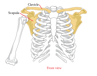
The scapula is also known as the shoulder blade. It is a flat, triangular bone located at the back of the shoulder. It connects the humerus (upper arm bone) with the clavicle (collarbone) and provides the foundation for the shoulder joint. This bone might seem small depending on where it was gotten from, but it is actually powerful when it comes to our ability to move our arms and shoulders.
So, why is the scapula so important? Well, it serves as the attachment point for many muscles involved in shoulder movement. These muscles allow us to reach, lift, pull, push, and rotate our arms in a wide range of motions. Without a properly functioning scapula, our shoulders would be limited in their movement and we would struggle to perform even the most basic daily tasks.
In addition to those roles above, the scapula also plays a key role in stabilising the shoulder joint. It helps to keep the humerus in place and prevent excessive motion that could lead to injury. Without this stability, our shoulders would be prone to dislocation and other issues that can seriously impact our ability to use our arms effectively.
The Scapula
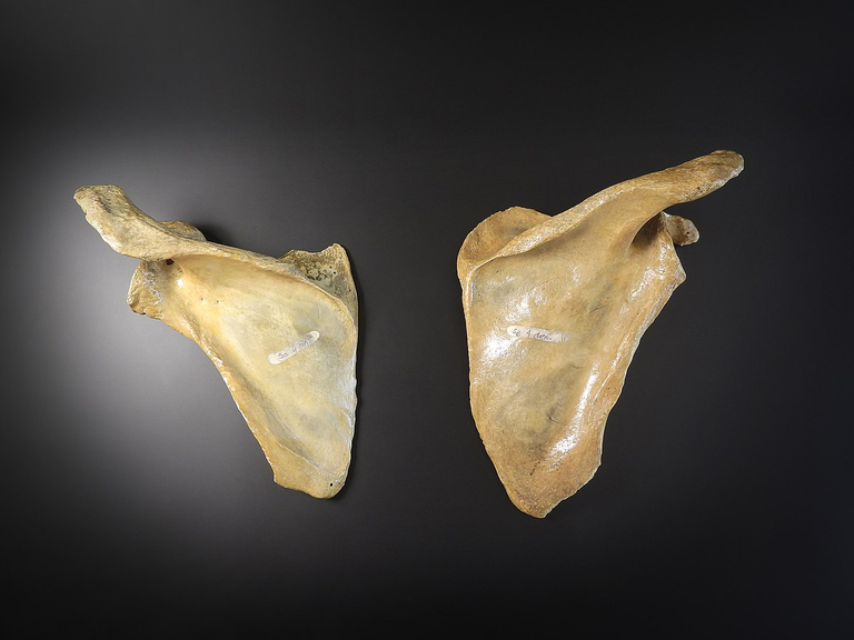
Source : Wikipedia
The scapula, also known as the shoulder blade, is a flat and triangular bone that lies on the upper part of your back. It is one of the three bones that form the shoulder joint, along with the clavicle (collarbone) and the humerus (upper arm bone). The scapula is a very important bone for your upper limb, as it provides attachment sites for many muscles that are involved in the movement and stabilization of the shoulder. In this article, we will describe the gross anatomy of the scapula, focusing on two aspects: its location and general shape, and its landmarks and features.
Location and General Shape
The scapula is located on the posterior aspect of the thorax, between the second and seventh ribs. It is slightly tilted forward, so that its anterior surface faces the ribs, and its posterior surface faces the skin. The scapula has three borders: the superior, medial, and lateral borders. The superior border is the shortest and thinnest, and it runs from the base of the coracoid process to the medial angle of the scapula. The medial border is the longest and thickest, and it runs from the medial angle to the inferior angle of the scapula. The lateral border is the most prominent, and it runs from the inferior angle to the glenoid cavity, where it articulates with the humerus.
The scapula also has three angles: the superior, inferior, and lateral angles. The superior angle is formed by the junction of the superior and medial borders, and it lies at the level of the second rib. The inferior angle is formed by the junction of the medial and lateral borders, and it lies at the level of the seventh rib. The lateral angle is the most important, as it bears the glenoid cavity, which is a shallow and oval depression that receives the head of the humerus.
The scapula has a general triangular shape, with a slightly concave anterior surface and a convex posterior surface. The anterior surface is divided into two parts by the subscapular fossa, which is a large and smooth area that occupies most of the surface. The posterior surface is divided into two parts by the spine of the scapula, which is a prominent and ridge-like projection that runs across the surface.
Landmarks and Features
The scapula has several landmarks and features that are important for the attachment of muscles and ligaments, as well as for the articulation with other bones. These landmarks and features are:
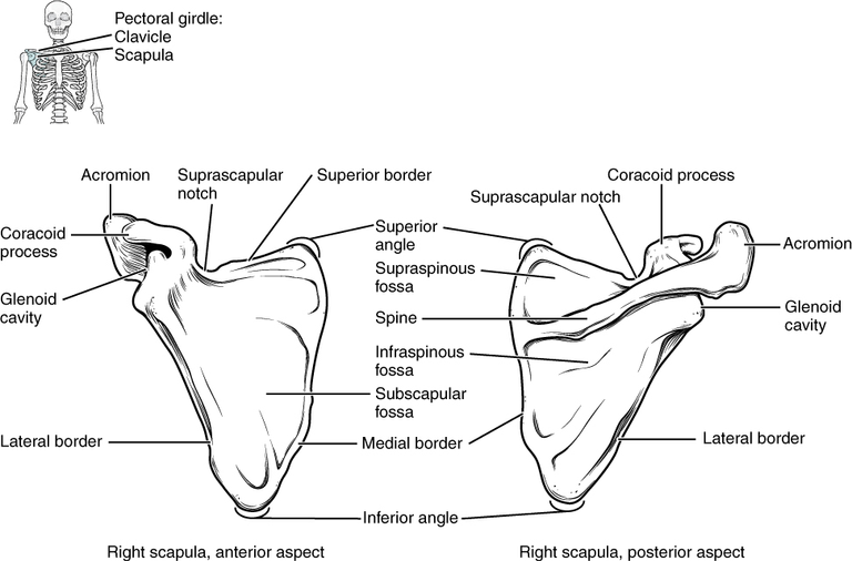
- The spine of the scapula, which is a prominent and ridge-like projection that runs across the posterior surface of the scapula. It divides the surface into two parts: the supraspinous fossa and the infraspinous fossa. The spine of the scapula ends laterally in the acromion process, which is a flat and broad projection that overhangs the glenoid cavity. The acromion process articulates with the clavicle at the acromioclavicular joint, and it provides attachment for the trapezius and deltoid muscles.
- The glenoid cavity, which is a shallow and oval depression that lies on the lateral angle of the scapula. It articulates with the head of the humerus at the glenohumeral joint, which is the main joint of the shoulder. The glenoid cavity is surrounded by a fibrocartilaginous rim called the glenoid labrum, which deepens the cavity and enhances the stability of the joint. The glenoid cavity also has a small tubercle on its superior part, called the supraglenoid tubercle, which provides attachment for the long head of the biceps brachii muscle.
- The coracoid process, which is a hook-like projection that arises from the superior border of the scapula, near the base of the neck. It curves anteriorly and laterally, and it lies below the clavicle. The coracoid process provides attachment for several muscles and ligaments, such as the pectoralis minor, the coracobrachialis, the short head of the biceps brachii, the coracoclavicular ligament, and the coracoacromial ligament.
- The subscapular fossa, which is a large and smooth area that occupies most of the anterior surface of the scapula. It provides attachment for the subscapularis muscle, which is one of the four muscles that form the rotator cuff, a group of muscles and tendons that stabilize the shoulder joint.
- The supraspinous fossa, which is a smaller and concave area that lies above the spine of the scapula, on the posterior surface. It provides attachment for the supraspinatus muscle, which is another muscle of the rotator cuff.
- The infraspinous fossa, which is a larger and convex area that lies below the spine of the scapula, on the posterior surface. It provides attachment for the infraspinatus muscle, which is also a muscle of the rotator cuff.
At this point , we have seen that the scapula is a very important bone for our shoulder, as it connects, supports, and moves it in various ways. The scapula also gives shape and definition to your upper back, and it can be easily felt and seen under your skin.
How The Shoulder Moves: Articulations and Muscles
The shoulder is one of the most mobile and versatile joints in our body. It allows us to perform a wide range of movements, such as lifting, throwing, reaching, and swinging. But how does the shoulder work? What are the bones, joints, and muscles that make up the shoulder? In this section, we will answer these questions and more, by describing the articulations and movements of the shoulder, and the muscles involved in scapular movements.
Glenohumeral Joint
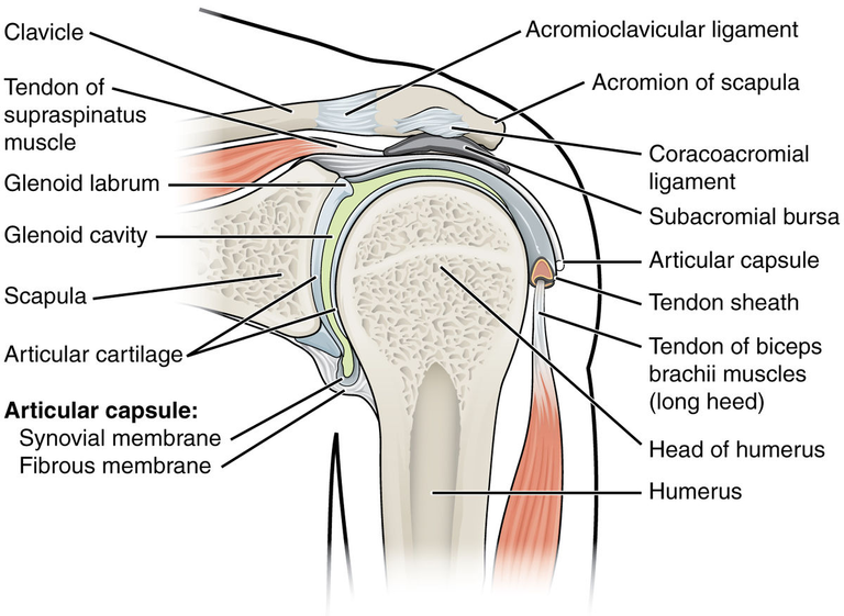
The glenohumeral joint is the main joint of the shoulder, and it is formed by the articulation of the head of the humerus (upper arm bone) and the glenoid cavity of the scapula (shoulder blade). The glenohumeral joint is a ball-and-socket joint, meaning that it has a spherical head that fits into a cup-like socket. This type of joint allows for a great degree of freedom of movement, but it also makes the joint less stable and more prone to injury.
The glenohumeral joint is surrounded by a fibrous capsule that encloses a synovial membrane that secretes synovial fluid, which lubricates the joint and reduces friction. The capsule is reinforced by several ligaments, such as the coracohumeral ligament, the glenohumeral ligaments, and the transverse humeral ligament. The capsule is also supported by a fibrocartilaginous rim called the glenoid labrum, which deepens the socket and increases the contact area between the head and the cavity.
The glenohumeral joint allows for six types of movements: flexion and extension, abduction and adduction, and medial and lateral rotation of the humerus. Flexion is the movement of the arm forward and upward, while extension is the movement of the arm backward and downward. Abduction is the movement of the arm away from the body, while adduction is the movement of the arm toward the body. Medial rotation is the movement of the arm inward, while lateral rotation is the movement of the arm outward. The glenohumeral joint also allows for a combination of these movements, called circumduction, which is the movement of the arm in a circular motion.
The glenohumeral joint is controlled by several muscles that originate from the scapula, the clavicle (collarbone), the sternum (breastbone), and the ribs, and insert on the humerus. These muscles are:
- The rotator cuff, which is a group of four muscles and tendons that surround the glenohumeral joint and stabilize it. The rotator cuff muscles are the supraspinatus, which abducts the humerus; the infraspinatus, which laterally rotates the humerus; the teres minor, which also laterally rotates the humerus; and the subscapularis, which medially rotates the humerus.
- The deltoid, which is a thick, triangular muscle that covers the shoulder. The deltoid has three parts: the anterior, middle, and posterior fibers. The anterior fibers flex and medially rotate the humerus; the middle fibers abduct the humerus; and the posterior fibers extend and laterally rotate the humerus.
- The pectoralis major, which is a large, fan-shaped muscle that covers the chest. The pectoralis major has two heads: the clavicular head and the sternal head. The clavicular head flexes, adducts, and medially rotates the humerus; the sternal head adducts and medially rotates the humerus.
- The latissimus dorsi, which is a broad, flat muscle that covers the lower back. The latissimus dorsi extends, adducts, and medially rotates the humerus.
- The teres major, which is a small, round muscle that lies below the teres minor. The teres major extends, adducts, and medially rotates the humerus.
- The coracobrachialis, which is a small, cylindrical muscle that lies under the coracoid process of the scapula. The coracobrachialis flexes and adducts the humerus.
- The biceps brachii, which is a large muscle that runs along the front of the upper arm. The biceps brachii has two heads: the long head and the short head. The long head originates from the supraglenoid tubercle of the scapula, and the short head originates from the coracoid process of the scapula. Both heads insert on the radial tuberosity of the radius (forearm bone). The biceps brachii flexes and supinates the forearm, as well as flexes the humerus.
- The triceps brachii, which is a large muscle that runs along the back of the upper arm. The triceps brachii has three heads: the long head, the lateral head, and the medial head. The long head originates from the infraglenoid tubercle of the scapula, and the lateral and medial heads originate from the posterior surface of the humerus. All three heads insert on the olecranon of the ulna (forearm bone). The triceps brachii extends and adducts the forearm, as well as extends the humerus.
Acromioclavicular Joint
The acromioclavicular joint is the joint between the acromion process of the scapula and the lateral end of the clavicle. The acromioclavicular joint is a plane joint, meaning that it has flat surfaces that allow for gliding movements. The acromioclavicular joint is surrounded by a fibrous capsule that encloses a synovial membrane that secretes synovial fluid. The capsule is reinforced by several ligaments, such as the acromioclavicular ligament, the coracoclavicular ligament, and the coracoacromial ligament.
The acromioclavicular joint allows for some movement of the scapula on the clavicle, such as upward and downward rotation, anterior and posterior tilting, and internal and external rotation. Upward rotation is the movement of the scapula upward and laterally, while downward rotation is the movement of the scapula downward and medially. Anterior tilting is the movement of the scapula forward, while posterior tilting is the movement of the scapula backward. Internal rotation is the movement of the scapula inward, while external rotation is the movement of the scapula outward.
The acromioclavicular joint is controlled by several muscles that originate from the scapula, the clavicle, the sternum, the ribs, and the spine, and insert on the scapula. These muscles are:
- The trapezius, which is a large, triangular muscle that covers the back of the neck and the upper part of the back. The trapezius has three parts: the upper, middle, and lower fibers. The upper fibers elevate and upwardly rotate the scapula; the middle fibers retract the scapula; and the lower fibers depress and upwardly rotate the scapula.
- The serratus anterior, which is a large, fan-shaped muscle that lies on the lateral aspect of the chest. The serratus anterior originates from the upper eight or nine ribs, and inserts on the medial border of the scapula. The serratus anterior protracts and upwardly rotates the scapula, as well as stabilizes it against the thoracic wall.
- The rhomboids, which are two muscles that lie deep to the trapezius. The rhomboids have two parts: the rhomboid major and the rhomboid minor. The rhomboid major originates from the spinous processes of the second to fifth thoracic vertebrae, and inserts on the medial border of the scapula, below the spine. The rhomboid minor originates from the spinous processes of the seventh cervical and first thoracic vertebrae, and inserts on the medial border of the scapula, at the base of the spine. The rhomboids retract and downwardly rotate the scapula, as well as elevate it.
- The levator scapulae, which is a thin, strap-like muscle that runs from the neck to the scapula. The levator scapulae originates from the transverse processes of the first to fourth cervical vertebrae, and inserts on the medial border of the scapula, above the spine. The levator scapulae elevates and downwardly rotates the scapula, as well as flexes and rotates the neck.
- The pectoralis minor, which is a small, triangular muscle that lies under the pectoralis major. The pectoralis minor originates from the third to fifth ribs, and inserts on the coracoid process of the scapula. The pectoralis minor depresses and anteriorly tilts the scapula, as well as elevates the ribs during inspiration.
Scapulothoracic Articulation
The scapulothoracic articulation is not a true joint, but rather a functional unit that consists of the scapula and the thoracic wall. The scapula glides over the posterior surface of the thoracic wall, forming a concave-convex relationship. The concave surface of the scapula is formed by the subscapular fossa, while the convex surface of the thoracic wall is formed by the ribs and the intercostal muscles. The scapulothoracic articulation is not stabilized by any ligaments or capsules, but rather by the muscles that attach the scapula to the thorax.
The scapulothoracic articulation allows for four types of movements: elevation and depression, protraction and retraction, upward and downward rotation, and anterior and posterior tilting of the scapula. Elevation is the movement of the scapula upward, while depression is the movement of the scapula downward. Protraction is the movement of the scapula forward, while retraction is the movement of the scapula backward. Upward rotation is the movement of the scapula upward and laterally, while downward rotation is the movement of the scapula downward and medially. Anterior tilting is the movement of the scapula forward, while posterior tilting is the movement of the scapula backward.
The scapulothoracic articulation is important for the movement and stability of the shoulder, as it allows the scapula to adjust its position and orientation according to the demands of the upper limb. The scapulothoracic articulation also affects the position of the glenoid cavity, which in turn affects the range of motion and stability of the glenohumeral joint.
Arterial Supply to the Scapula
The scapula receives blood from several arteries that branch off from the subclavian artery, which is a major artery that arises from the aortic arch and supplies the upper limb. The main arteries that supply the scapula are:
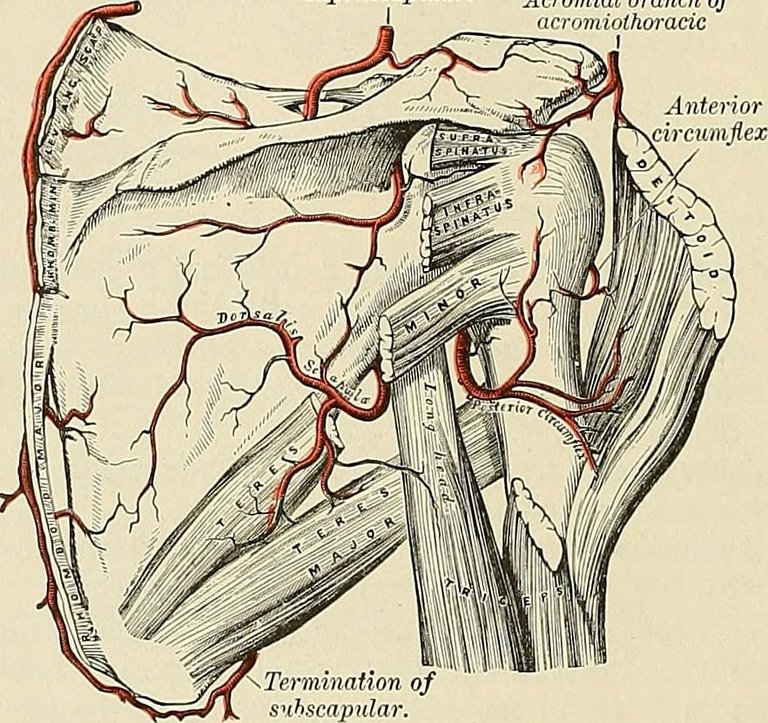
- The suprascapular artery, which is a branch of the thyrocervical trunk, which is a branch of the subclavian artery. The suprascapular artery runs along the superior border of the scapula, and passes through the suprascapular notch, which is a small opening on the scapula that allows the passage of the suprascapular nerve. The suprascapular artery supplies the supraspinatus and infraspinatus muscles, which are two of the four muscles that form the rotator cuff, a group of muscles and tendons that stabilize the shoulder joint.
- The dorsal scapular artery, which is a branch of the subclavian artery or the transverse cervical artery, which is a branch of the thyrocervical trunk. The dorsal scapular artery runs along the medial border of the scapula, and supplies the rhomboids and the levator scapulae muscles, which are involved in the retraction and elevation of the scapula, respectively.
- The circumflex scapular artery, which is a branch of the subscapular artery, which is a branch of the axillary artery, which is a continuation of the subclavian artery. The circumflex scapular artery runs along the lateral border of the scapula, and passes through the triangular space, which is a small gap between the teres minor, the teres major, and the long head of the triceps brachii muscles. The circumflex scapular artery supplies the teres minor, the teres major, and the subscapularis muscles, which are involved in the lateral rotation, extension, and medial rotation of the humerus, respectively.
- The thoracodorsal artery, which is a branch of the subscapular artery. The thoracodorsal artery runs along the inferior angle of the scapula, and supplies the latissimus dorsi muscle, which is involved in the extension, adduction, and medial rotation of the humerus.
The scapula receives blood from various sources, and distributes it to the muscles that attach to it and control its movements.
Venous Drainage
The scapula drains blood from several veins that correspond to the arteries that supply it. The main veins that drain the scapula are:
- The suprascapular vein, which is a tributary of the external jugular vein, which is a major vein that drains the head and neck. The suprascapular vein runs along the superior border of the scapula, and passes through the suprascapular notch. The suprascapular vein drains the supraspinatus and infraspinatus muscles.
- The dorsal scapular vein, which is a tributary of the subclavian vein, which is a major vein that drains the upper limb. The dorsal scapular vein runs along the medial border of the scapula, and drains the rhomboids and the levator scapulae muscles.
- The circumflex scapular vein, which is a tributary of the axillary vein, which is a continuation of the subclavian vein. The circumflex scapular vein runs along the lateral border of the scapula, and passes through the triangular space. The circumflex scapular vein drains the teres minor, the teres major, and the subscapularis muscles.
- The thoracodorsal vein, which is a tributary of the axillary vein. The thoracodorsal vein runs along the inferior angle of the scapula, and drains the latissimus dorsi muscle.
Nerve Supply to the Scapular Region
The scapula receives nerve supply from several nerves that branch off from the brachial plexus, which is a network of nerves that originates from the spinal nerves of the cervical and thoracic regions, and supplies the upper limb. The main nerves that supply the scapula are:
- The suprascapular nerve, which is a branch of the superior trunk of the brachial plexus, which is formed by the union of the fifth and sixth cervical spinal nerves. The suprascapular nerve runs along the superior border of the scapula, and passes through the suprascapular notch. The suprascapular nerve innervates the supraspinatus and infraspinatus muscles, and provides sensory innervation to the glenohumeral joint and the acromioclavicular joint.
- The dorsal scapular nerve, which is a branch of the fifth cervical spinal nerve, or sometimes the fourth or sixth cervical spinal nerves. The dorsal scapular nerve runs along the medial border of the scapula, and innervates the rhomboids and the levator scapulae muscles.
- The subscapular nerves, which are branches of the posterior cord of the brachial plexus, which is formed by the union of the posterior divisions of the upper, middle, and lower trunks of the brachial plexus. The subscapular nerves include the upper subscapular nerve, which innervates the subscapularis muscle; the lower subscapular nerve, which innervates the subscapularis and the teres major muscles; and the thoracodorsal nerve, which innervates the latissimus dorsi muscle.
- The axillary nerve, which is a branch of the posterior cord of the brachial plexus. The axillary nerve runs along the lateral border of the scapula, and passes through the quadrangular space, which is a small gap between the teres minor, the teres major, the long head of the triceps brachii, and the humerus. The axillary nerve innervates the deltoid and the teres minor muscles, and provides sensory innervation to the skin over the deltoid and the shoulder joint.
The scapula receives nerve supply from various sources, and distributes it to the muscles that attach to it and control its movements, as well as to the skin and the joints of the shoulder.
Formation of the Scapula During Embryonic Development
The scapula is derived from the mesoderm, which is one of the three primary germ layers that form during the early development of the embryo. The mesoderm gives rise to various tissues and organs, such as the muscles, the bones, the blood vessels, and the heart. The mesoderm is divided into several regions, such as the paraxial mesoderm, the intermediate mesoderm, and the lateral plate mesoderm.
The scapula is formed from the lateral plate mesoderm, which is the most lateral region of the mesoderm. The lateral plate mesoderm splits into two layers: the somatic mesoderm, which is adjacent to the ectoderm (the outermost layer of the embryo), and the splanchnic mesoderm, which is adjacent to the endoderm (the innermost layer of the embryo). The somatic mesoderm forms the somatopleure, which gives rise to the body wall and the limbs. The splanchnic mesoderm forms the splanchnopleure, which gives rise to the visceral organs and the blood vessels.
The scapula is formed from the somatopleure of the lateral plate mesoderm, specifically from the region that corresponds to the upper limb bud. The upper limb bud is a small outgrowth of the somatopleure that appears around the fourth week of embryonic development, on the lateral side of the trunk. The upper limb bud consists of a core of mesenchymal cells, which are undifferentiated cells that can give rise to various types of cells, such as the bone cells and the muscle cells. The upper limb bud is covered by a layer of ectodermal cells, which form the apical ectodermal ridge (AER), a thickened structure that induces the growth and patterning of the limb.
The scapula is formed from the proximal part of the upper limb bud, which is the part that is closest to the trunk. The proximal part of the upper limb bud forms the shoulder girdle, which consists of the scapula and the clavicle. The scapula is formed from the dorsal part of the proximal limb bud, which is the part that faces the back. The dorsal part of the proximal limb bud forms a cartilaginous plate, which is a flat and flexible structure that is composed of cartilage cells. The cartilaginous plate is the precursor of the scapula, and it undergoes a series of changes to become the mature bone.
Ossification Process
The scapula undergoes endochondral ossification, which is a process that involves the replacement of the cartilage by bone. Endochondral ossification occurs in most of the bones of the body, such as the long bones and the vertebrae. Endochondral ossification involves several steps, such as the formation of a primary ossification center, the formation of a secondary ossification center, and the formation of an epiphyseal plate.
The scapula has two primary ossification centers, which are the sites where the bone formation begins. The first primary ossification center appears around the eighth week of embryonic development, in the coracoid process of the scapula. The coracoid process is a hook-like projection that arises from the superior border of the scapula, near the base of the neck. The second primary ossification center appears around the twelfth week of embryonic development, in the body of the scapula. The body of the scapula is the main part of the bone, and it has a triangular shape.
The scapula has several secondary ossification centers, which are the sites where the bone formation continues after birth. The secondary ossification centers appear around puberty, and they are located in the acromion process, the glenoid cavity, the inferior angle, and the medial border of the scapula. The acromion process is a flat and broad projection that overhangs the glenoid cavity, and it articulates with the clavicle at the acromioclavicular joint. The glenoid cavity is a shallow and oval depression that lies on the lateral angle of the scapula, and it articulates with the head of the humerus at the glenohumeral joint. The inferior angle is formed by the junction of the medial and lateral borders of the scapula, and it lies at the level of the seventh rib. The medial border is the longest and thickest border of the scapula, and it runs from the medial angle to the superior angle of the scapula.
The scapula does not have an epiphyseal plate, which is a layer of cartilage that separates the primary and secondary ossification centers, and allows for the growth of the bone. The scapula grows by appositional growth, which is a process that involves the addition of new bone tissue on the surface of the existing bone. The scapula reaches its adult size and shape around the age of 25, when the secondary ossification centers fuse with the primary ossification centers.
Variations in Scapular Anatomy
The scapula is a very variable bone, and it can show some differences in its size, shape, and features. Some of the common variations in scapular anatomy are:
- The os acromiale, which is a condition where the acromion process remains unfused with the rest of the scapula, and forms a separate bone. The os acromiale occurs in about 8% of the population, and it is more common in males than in females. The os acromiale can cause some problems, such as shoulder pain, impingement syndrome, and rotator cuff tears.
- The suprascapular notch, which is a small opening on the superior border of the scapula, that allows the passage of the suprascapular nerve. The suprascapular notch can vary in its shape and size, and it can be classified into six types: type I, which is a V-shaped notch; type II, which is a U-shaped notch; type III, which is a wide U-shaped notch; type IV, which is a narrow U-shaped notch; type V, which is a foramen (a complete hole); and type VI, which is an absent notch. The suprascapular notch can also be bridged by a ligament or a bone, which can compress the suprascapular nerve and cause nerve injury.
- The infraglenoid tubercle, which is a small projection on the inferior part of the glenoid cavity, that provides attachment for the long head of the triceps brachii muscle. The infraglenoid tubercle can vary in its prominence and shape, and it can be classified into four types: type A, which is a flat tubercle; type B, which is a round tubercle; type C, which is a hook-shaped tubercle; and type D, which is a bifid (split) tubercle. The infraglenoid tubercle can also be absent in some cases.
As you can see, the scapula is a very complex and variable bone, and it can show some differences in its anatomy. These differences can affect the function and health of the shoulder, and they can be detected by various methods, such as X-rays, CT scans, MRI scans, and physical examination.
This is where I will drop the pen on this topic. Next, we'll be looking at another bone of the upper limb which is the humerus.
I hope this article helps you understand more
References
(1) Scapula: Anatomy and clinical notes | Kenhub. https://www.kenhub.com/en/library/anatomy/scapula.
(2) Scapula (Shoulder Blade) – Anatomy, Location, & Labeled Diagram. https://www.theskeletalsystem.net/arm-bones/scapula.html.
(3) Scapula: Anatomy and clinical notes | Kenhub. https://www.kenhub.com/en/library/anatomy/scapula.
(4) Scapula | Radiology Reference Article | Radiopaedia.org. https://radiopaedia.org/articles/scapula.
(5) Scapula | Skeleton of the upper limb | Upper Extremity | Anatomy.app .... https://anatomy.app/article/skeleton-of-the-upper-limb/scapula.
(6) Scapula: Anatomy and clinical notes | Kenhub. https://www.kenhub.com/en/library/anatomy/scapula.
(7) Circumflex scapular artery: Anatomy, branches, supply | Kenhub. https://www.kenhub.com/en/library/anatomy/circumflex-scapular-artery.
(8) Arterial Anastomosis around Scapula (Scapular Anastomosis). https://www.earthslab.com/anatomy/arterial-anastomosis-around-scapula-scapular-anastomosis/.
(9) Scapula: Anatomy and clinical notes | Kenhub. https://www.kenhub.com/en/library/anatomy/scapula.
(10) Scapular anastomosis - Wikipedia. https://en.wikipedia.org/wiki/Scapular_anastomosis.
(11) Scapula | Complete Anatomy - Elsevier. https://www.elsevier.com/resources/anatomy/skeletal-system/appendicular-skeleton/scapula/23344.
(12) The clavicle and scapula | Acland's Video Atlas of Human Anatomy. https://aclandanatomy.com/multimediaplayer.aspx?multimediaid=10528033.
(13) Scapula bone: anatomy, structure and labeled diagram - GetBodySmart. https://www.getbodysmart.com/upper-limb-bones/scapula-anterior/.
(14) Dorsal scapular nerve: Origin, course and function | Kenhub. https://www.kenhub.com/en/library/anatomy/dorsal-scapular-nerve.
(15) Dorsal Scapular Nerve - Course - Motor Functions - TeachMeAnatomy. https://teachmeanatomy.info/encyclopaedia/d/dorsal-scapular-nerve/.
(16) Dorsal scapular nerve - Wikipedia. https://en.wikipedia.org/wiki/Dorsal_scapular_nerve.
(17) The Intrinsic Muscles of the Shoulder - TeachMeAnatomy. https://teachmeanatomy.info/upper-limb/muscles/shoulder/intrinsic/.
(18) Anatomy of the Scapula | SpringerLink. https://link.springer.com/chapter/10.1007/978-3-319-53584-5_1.
(19) Anatomy, Back, Scapula - Abstract - Europe PMC. https://europepmc.org/article/NBK/nbk531475.
(20) Scapula: Anatomy, Function, and Treatment - Verywell Health. https://www.verywellhealth.com/scapula-anatomy-4682581.


I am a complete beginner who resides in Africa's Western Hemisphere. My name is James, but you may reach out to me through the Facebook page [James Kossy] (https://www.facebook.com/christ.messenger.904) Physics, chemistry, and biology are the three topics that I find most enjoyable. My current studies are taking place at the university level, with the intention of becoming a recognized professional in physiotherapy. I am fascinated by all things technological, and I take pleasure in contributing to the fascinating technological advancements that are taking place throughout the world today. In my spare time, I'd like to learn more about programming and help others with any technical problems they may be having. 💞 ***🌹❤️ Thank you so much to everyone who has supported me thus far. ****💞 At the moment, I don't have the right words to say how much I appreciate all of your help. You never cease to astonish me with your generosity. For me, this has turned into a haven of enjoyment. Thanks to colleagues like you, this has all been possible. You've been a great support for me. Everything you have done for me and my family has been greatly appreciated, and I will always be grateful to you. 💕.
Posted Using InLeo Alpha
Articulation of clavicle with the flat triangular scapula is quite interesting. Thought I had read about a cavity which articulates with the head of the humerus to form the shoulder joint. Can't remember the name now.
Glenoid cavity of the lateral angle of the scapula lodges the head of the humerus to form the glenohumeral joint. That's the name
Yes. Now I remember
I will always say that every part of the body is important because while you might not take note of the job that part of the body does because it is not as visible as that of the heart of the brain, it is actually working, and the shoulder bone is one of those body parts.
Yes, you're right and thanks for stopping by
Thanks for your contribution to the STEMsocial community. Feel free to join us on discord to get to know the rest of us!
Please consider delegating to the @stemsocial account (85% of the curation rewards are returned).
You may also include @stemsocial as a beneficiary of the rewards of this post to get a stronger support.