En el post anterior hablamos sobre las lesiones pulpares descritas en una serie de patología que definen la condicion de la pulpa. Con la ayuda de las técnicas y la metodología desarrollada en la endodoncia se caracteriza las diferentes lesiones y la severidad del daño en la pulpa.
In the previous post we talked about the pulp lesions described in a series of pathology that define the condition of the pulp. With the help of the techniques and methodology developed in endodontics we characterized the different lesions and the severity of the pulp damage.

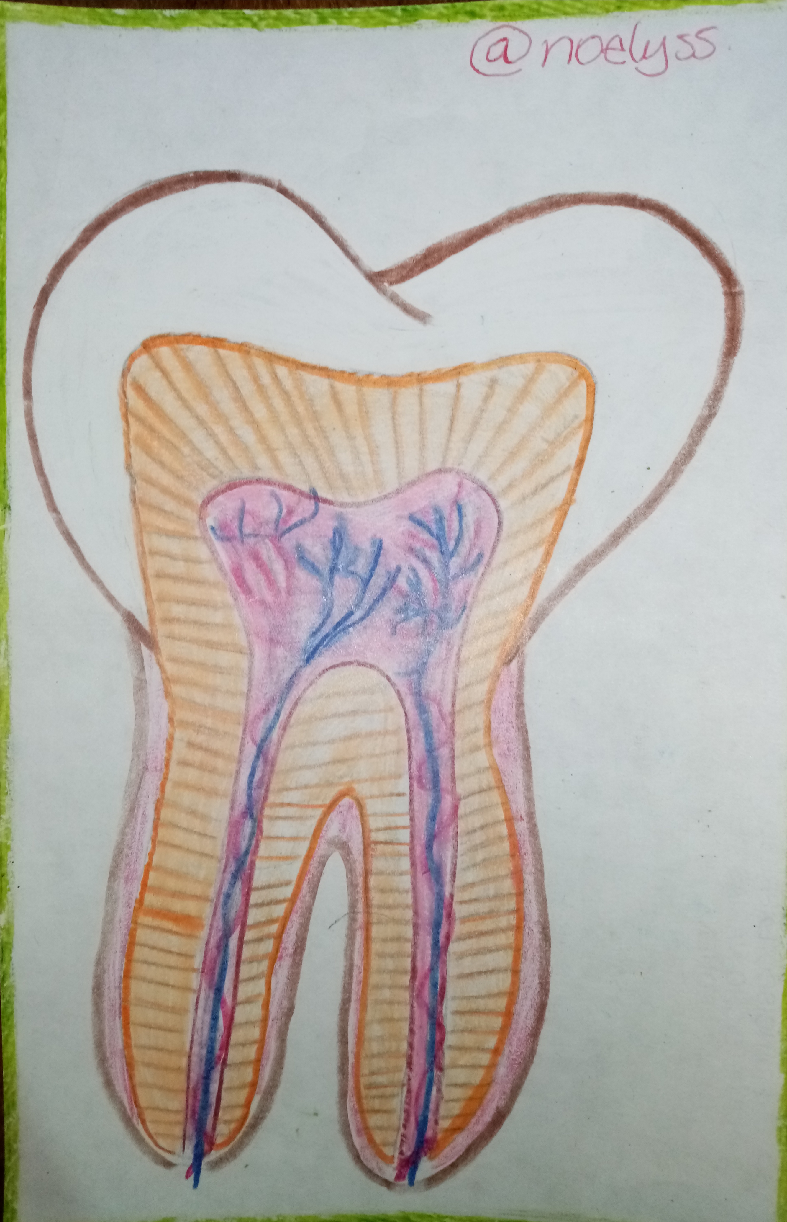
Patología pulpar | Pulpal Pathology
Además de las lesiones causadas por caries o otro tipo de infección existen casos donde la pulpa puede presentar deterioro propio de un desorden celular tales como
Resorcion dentinaria interna : Es un proceso en el cual las células mesenquimatosas1 inactivas se transforman en dentinoclastos2, es decir, se genera una expansión de la pulpa al punto de romper la dentina, de manera que se puede notar una inflamación o cierto traumatismos que hace ver a la pulpa más grande por reducción del tamaño de la dentina.
El fenómeno puede comenzar en la cámara pulpar o en el conducto radicular, lo cual puede llegar a perforar la raíz de adentro hacia afuera. No se sabe ' con exactitud a qué se debe esta anomalia, aunque afortunadamente es indolora y puede tratarse mediante un tratamiento de conducto.
In addition to lesions caused by caries or other types of infection there are cases where the pulp may present deterioration due to a cellular disorder such as
Internal dentin resorption: It is a process in which inactive mesenchymal cells1 are transformed into dentinoclasts2, that is, an expansion of the pulp is generated to the point of breaking the dentin, so that you can notice an inflammation or some trauma that makes the pulp look bigger by reducing the size of the dentin.
The phenomenon can begin in the pulp chamber or in the root canal, which can perforate the root from the inside out. It is not known exactly what causes this anomaly, although fortunately it is painless and can be treated by root canal treatment.
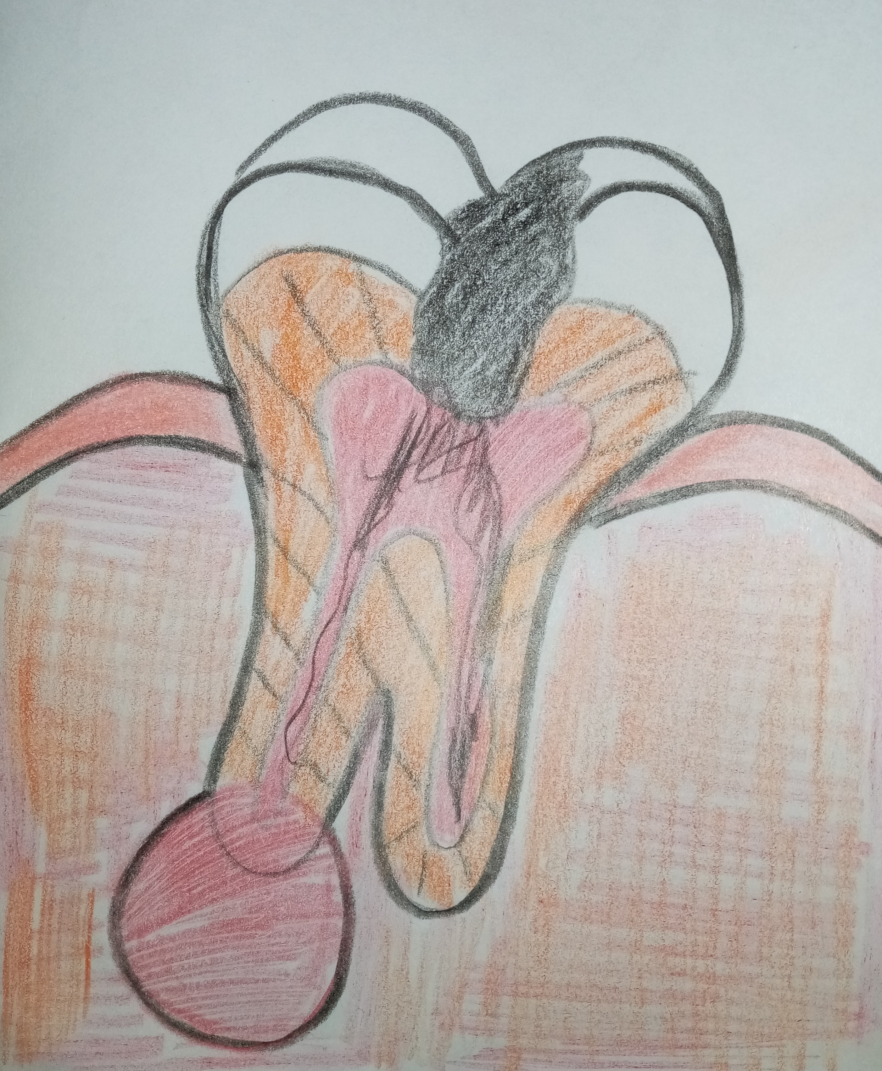
Metamorfosis calcica: Viene a ser lo contrario al fenómeno anterior, ya que trata de la acumulación de masa calcificada dentro del tejido pulpar, lo que reduce todos los elementos celulares como fibras y vasos sanguíneos y en general todo el espacio pulpar, además y por el exceso de dentina se puede apreciar una coloración amarillenta más extensa.
Más específicamente sucede que los tubulos dentinarios se llenan de depósitos minerales tales como cristales de hidroxiapatita 3 impidiendo que los irritantes se difundan a través de los tubos. Con respecto a las pruebas no se tinen respuesta a la percusión y la palpación, tal anómalia no muestra sensibilidad en el ligamento periodontal.
Calcific metamorphosis: It is the opposite of the previous phenomenon, since it is the accumulation of calcified mass within the pulp tissue, which reduces all cellular elements such as fibers and blood vessels and in general all the pulp space, in addition and due to the excess of dentin, a more extensive yellowish coloration can be seen.
More specifically it happens that the dentinal tubules are filled with mineral deposits such as hydroxyapatite 3 crystals preventing the irritants to diffuse through the tubes. With respect to testing, there is no response to percussion and palpation, such abnormality does not show sensitivity in the periodontal ligament.
Por otro lado también podríamos ver cambios pulpares regresivos cuyas alteraciones no son infecciosas y puede deberse a factores casuales como traumatismo, preparación de cavidades, inflamaciones periodónticas, etc.
On the other hand, we could also see "regressive pulp changes" whose alterations are not infectious and may be due to casual factors such as trauma, cavity preparation, periodontal inflammations, etc.
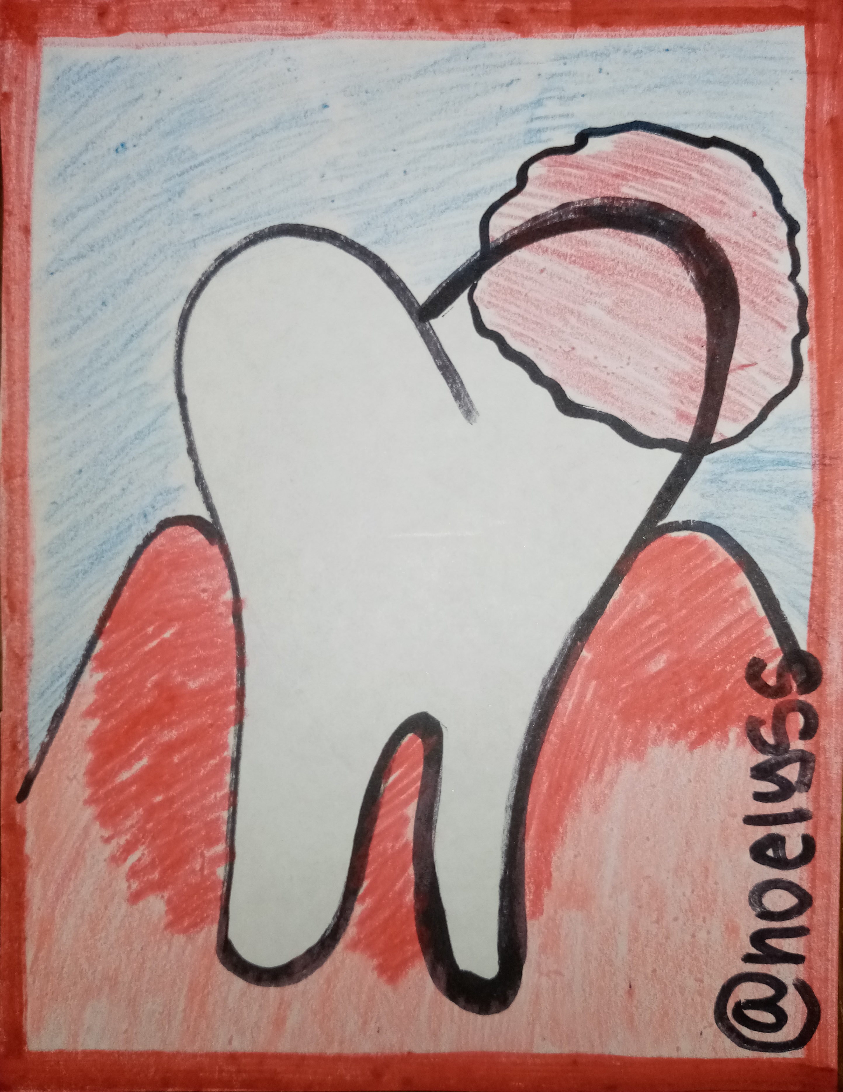
Patalogía periapical | Periapical Pathology
En la parte inferior del diente nos encontramos con daños denominados patologías periapicales, es decir, los problemas que se presentan alrededor de la raíz o ápice del diente. Dentro de estas enfermedades están la periodontitis y los abscesos con una característica en común y es la presencia de dolor a la percusión y la palpación.
Periodontitis apical aguda: es un proceso inflamatorio agudo alrededor del ápice, como consecuencia de una infección pulpar o traumatismo por la mala manipulación de instrumentos en los conductos radiculares. El dolor presente es intenso con una respuesta rápida a la percusión, inflamación del ligamento periodontal y posible movilidad.
In the lower part of the tooth we find damage called periapical pathologies, that is, problems that occur around the root or apex of the tooth. Among these diseases are periodontitis and abscesses with one characteristic in common and that is the presence of pain on percussion and palpation.
Acute apical periodontitis: is an acute inflammatory process around the apex, as a consequence of pulp infection or trauma due to bad manipulation of instruments in the root canals. The pain present is intense with a rapid response to percussion, inflammation of the periodontal ligament and possible mobility.
Periodontitis apical crónica: es una lesion periapical asintomática y crónica que generalmente viene acompañada de una resorción osea periradicular la cual es visible en las radiografías. La destrucción del periodonto apical es consecuencia de una necrosis pulpar, por tanto las pruebas de vitalidad pulpar son negativas aunque puede haber sensibilidad a la palpación.
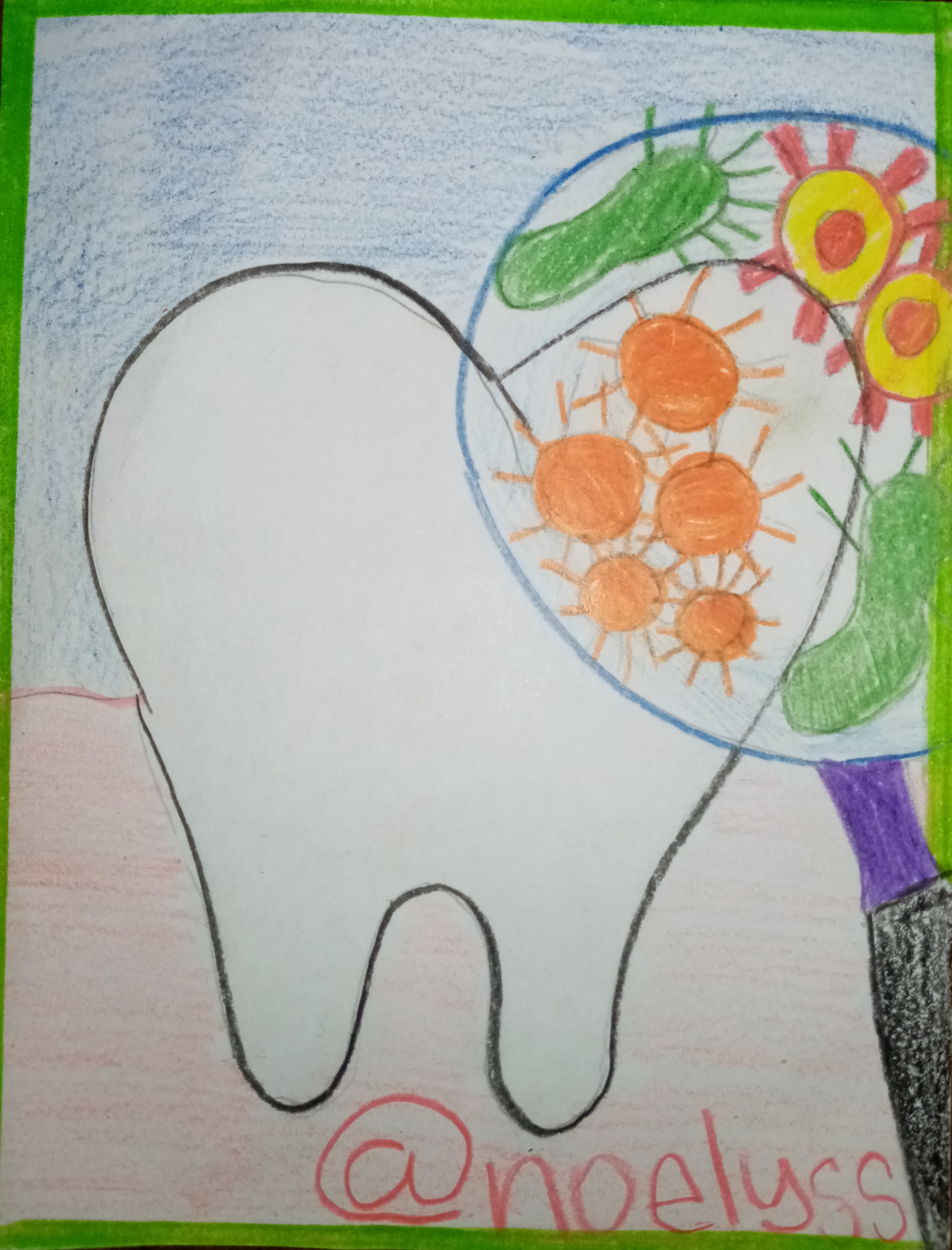
Absceso periapical agudo: es la colección de pus iniciada a nivel de los tejidos periapicales de un diente. Se tiene un dolor moderado o fuerte a la percusión y la palpación, también puede haber aumento de movilidad, en casos avanzados puede haber fiebre, malestar general y leucocitos, el fondo del vestibulo y los tejidos faciales próximo al diente están inflamados.
Absceso periapical crónico: consiste en la formación de un exudado periapical purulento, es decír, una colección de pus con drenaje espontáneo hacia el exterior a través de un trayecto fistuloso. El dolor se manifiesta se forma, desaparece al drenar porque lo que pasa como una lesión asintomática.
Chronic apical periodontitis is an asymptomatic and chronic periapical lesion that is usually accompanied by periradicular bone resorption which is visible on radiographs. The destruction of the apical periodontium is a consequence of pulp necrosis, therefore pulp vitality tests are negative although there may be sensitivity to palpation.
Acute periapical abscess: is the collection of pus initiated at the level of the periapical tissues of a tooth. There is moderate or severe pain on percussion and palpation, there may also be increased mobility, in advanced cases there may be fever, malaise and leukocytes, the vestibular fundus and facial tissues near the tooth are inflamed.
Chronic periapical abscess: consists of the formation of a purulent periapical exudate, i.e., a collection of pus with spontaneous drainage to the outside through a fistulous tract. The pain is manifested is formed, disappears when drained because it passes as an asymptomatic lesion.
Últimos comentarios| Last comments
Cómo pueden observar en un caso agudo el dolor es rápido e intenso, mientras que el crónico toma más tiempo en desarrollarse y tiende a ser asintomático, ambas lesiones se producen por necrosis por lo que las prueba de vitalidad son negativas.
Las lesiones pulpares y periapicales constituyen uno de los problemas más comunes en la cavidad bucal del paciente, aunque no determinan un registro absoluto de enfermedades, el conocimiento recopilado en este tema nace de la experiencia así como de la investigación lo cual ofrece una preparación adecuada que nos ayuda a identificar y diagnosticar la severidad de una lesión, de aquí que iniciar un plan que de solución al problema resulte adecuado y efectivo.
As you can observe in an acute case the pain is fast and intense, while the chronic one takes more time to develop and tends to be asymptomatic, both lesions are produced by necrosis so the vitality tests are negative.
Pulpal and periapical lesions are one of the most common problems in the oral cavity of the patient, although they do not determine an absolute record of disease, the knowledge gathered on this subject is born of experience and research which provides adequate preparation that helps us to identify and diagnose the severity of a lesion, hence initiate a plan to solve the problem is appropriate and effective.
Ha sido un placer compartir con ustedes este contenido, el cual iré expandiendo según la disponibilidad me lo permita, espero les sea de ayuda e interes y no duden en preguntar cual duda.
It has been a pleasure to share with you this content, which I will expand as availability allows me, I hope it will be of help and interest and do not hesitate to ask any questions.
 créditos @doze


Texto traducido en Deelp
Congratulations @noelyss! You have completed the following achievement on the Hive blockchain and have been rewarded with new badge(s):
Your next target is to reach 14000 upvotes.
You can view your badges on your board and compare yourself to others in the Ranking
If you no longer want to receive notifications, reply to this comment with the word
STOPTo support your work, I also upvoted your post!
Check out the last post from @hivebuzz:
Support the HiveBuzz project. Vote for our proposal!
Does calcific metamorphosis is more hard than the teeth or mineralization is poor which makes it weak ?
Hi @medical-hall thank you so much for reading and asking. With regard to calcium deposits certainly weaken the tooth as it eliminates irrigation and therefore vitality, which makes the tooth prone to produce