Treatment of diseases such as cancer often requires the placement of intravenous catheters for the long-term delivery of medications through the bloodstream. The catheters stay in the vein between chemotherapy sessions and are rinsed with anticoagulant solutions after each use. Unfortunately, the catheters and the solutions used to rinse them can be a source of danger, rather than a critical part of the treatment plan. Such was the case in December 2004 when an anticoagulant solution contaminated with the bacterium Pseudomonas fluorescens was used to rinse the intravenous catheters of patients. Thirty-six patients in four different states developed blood infections from the contaminated solution. The Centers for Disease Control and Prevention (CDC) investigated the outbreak, and by the end of January 2005, the U.S. Food and Drug Administration had alerted the nation and all existing bottles of contaminated solution were recalled. Problem solved? Unfortunately not.
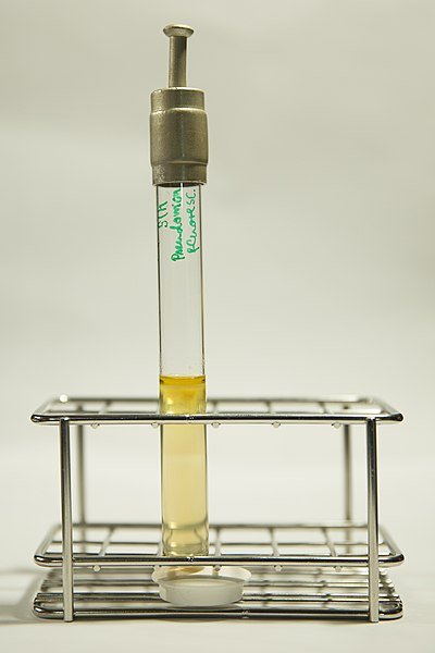
Image Source: Wikimedia CC BY-SA 4.0
In March 2005 the CDC was again alerted that 28 cancer patients developed bloodstream infections caused by P. fluorescens. Investigations determined that the infections were caused by the same P. fluorescens strain isolated from the cancer patients in 2004. These new patients had been exposed to the contaminated solution many months earlier but did not become ill. Thus they were not identified in the 2004 outbreak. Why was their illness so delayed? Subsequent CDC investigation showed that the catheters in the new patients had a P. fluorescens biofilm. The CDC hypothesized that the biofilm enabled the bacterium to reproduce, increasing its numbers from that in the original contaminating solution. Much after this initial exposure, the bacterial cells in the biofilm were dislodged when the catheters were flushed with sterile solutions. The dislodged cells were the cause of the blood infections. For these patients, physical forces released the cells from the biofilms, but it has long been known that bacteria have mechanisms for "deciding" when to establish a biofilm and when to "let go" and escape the biofilm. This involves sensing their environment and then altering cellular processes to make the switch. Switching back and forth between free-swimming (planktonic) and biofilm (sessile) growth is an example of a regulated behavior. Importantly, understanding how the change is made is critical to developing strategies for controlling biofilm formation and dissolution. A series of experiments culminating in a 2011 report have begun to reveal the mechanism for letting go, at least in one situation. The model includes an adhesion molecule located in the outer membrane (LapA), another protein (LapG), and describes how they work to control cells leaving a biofilm when phosphate levels are low.
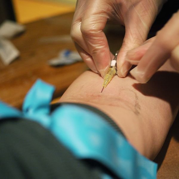
Image Source: Wikimedia CC BY-SA 2.0 Author: Thirteen Of Clubs from Minneapolis
Intravenous catheters such this are often used to deliver drugs. However, they can also serve as a portal of entry for pathogenic bacteria and as a site for biofilm formation.
Why phosphate? Phosphate is needed for synthesis of nucleic acids. No phosphate, no DNA replication or transcription, dead cell. When phosphate is abundant, P. f/uorescens cells want to stay put. They do so by sticking to a surface with LapA, the adhesion protein. Biofilm building then commences. However, when phosphate levels wane, if the cells are to survive, they must escape the biofilm and hunt for a location with more phosphate. They accomplish this with LapG, a periplasmic protease that cleaves LapA, removing it from the outer membrane. Without LapA holding them to the surface, the bacterial cells are free to leave the biofilm. The capacity of P. fluorescens to stay or leave a biofilm is just one example of the many"decisions" microbes make. Bacteria are constantly detecting changes in their environment and responding accordingly. Cells use two general approaches to regulate cellular processes. (1) They can alter the activity of enzymes and other proteins. This type of regulation occurs after the protein is synthesized and is called posttranslational control. (2) They can change the rate of synthesis of enzymes and other proteins. This type of regulation is often called regulation of gene expression. Posttranslational control acts rapidly to adjust metabolic activity and other processes from moment to moment. The control of gene expression occurs over longer intervals. For example, the Escherichia coli chromosome can code for about 4,500 polypeptides, yet only a fraction are synthesized at any one time. Regulation of gene expression conserves energy and raw materials, maintains the balance between the amounts of various cell proteins, and enables microbes to acclimate to long-term environmental change. Thus control of gene expression complements posttranslational control.
Thanks for reading this, comments and questions are entertained.
References
A Class Lecture, MCB303 (Immunology) 😁😁
WARNING - The message you received from @sadiajafri is a CONFIRMED SCAM!
DO NOT FOLLOW any instruction and DO NOT CLICK on any link in the comment!
For more information, read this post:
https://steemit.com/steemit/@arcange/phishing-site-reported-postupper-dot-ml
If you find my work to protect you and the community valuable, please consider to upvote this warning or to vote for my witness.